2ALZ

Ternary Complex of hPoli with DNA and dCTP


Experimental Data Snapshot

  • Method: X-RAY DIFFRACTION
  • Resolution: 2.50 Å
  • R-Value Free: 0.272 
  • R-Value Work: 0.228 

wwPDB Validation   3D Report Full Report


Ligand Structure Quality Assessment 


This is version 1.3 of the entry. See complete history


Literature

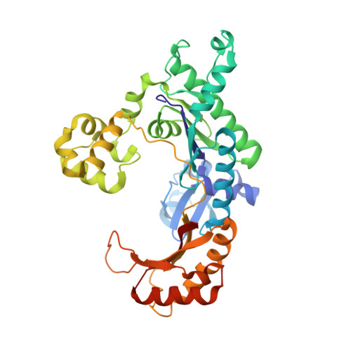
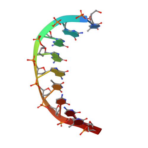
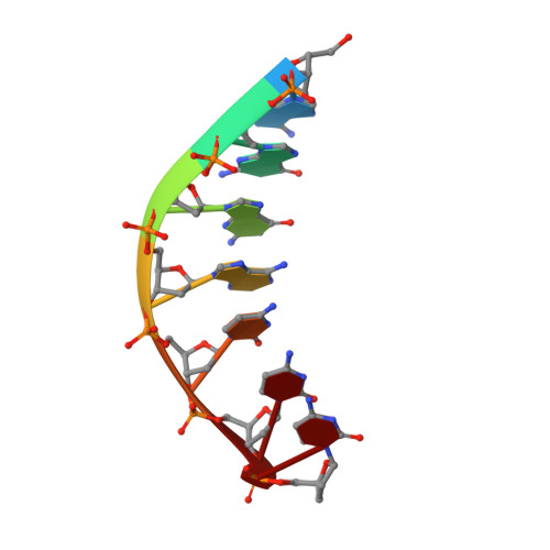
Human DNA Polymerase iota Incorporates dCTP Opposite Template G via a G.C+ Hoogsteen Base Pair.

Nair, D.T.Johnson, R.E.Prakash, L.Prakash, S.Aggarwal, A.K.

(2005) Structure 13: 1569-1577

  • DOI: https://doi.org/10.1016/j.str.2005.08.010
  • Primary Citation of Related Structures:  
    2ALZ

  • PubMed Abstract: 

    Human DNA polymerase iota (hPoliota), a member of the Y family of DNA polymerases, differs in remarkable ways from other DNA polymerases, incorporating correct nucleotides opposite template purines with a much higher efficiency and fidelity than opposite template pyrimidines. We present here the crystal structure of hPoliota bound to template G and incoming dCTP, which reveals a G.C + Hoogsteen base pair in a DNA polymerase active site. We show that the hPoliota active site has evolved to favor Hoogsteen base pairing, wherein the template sugar is fixed in a cavity that reduces the C1'-C1' distance across the nascent base pair from approximately 10.5 A in other DNA polymerases to 8.6 A in hPoliota. The rotation of G from anti to syn is then largely in response to this curtailed C1'-C1' distance. A G.C+ Hoogsteen base pair suggests a specific mechanism for hPoliota's ability to bypass N(2)-adducted guanines that obstruct replication.


  • Organizational Affiliation

    Structural Biology Program, Department of Physiology and Biophysics, Mount Sinai School of Medicine, Box 1677, 1425 Madison Avenue, New York, New York 10029, USA.


Macromolecules

Find similar proteins by:  (by identity cutoff)  |  3D Structure
Entity ID: 3
MoleculeChains Sequence LengthOrganismDetailsImage
DNA polymerase iotaC [auth A]390Homo sapiensMutation(s): 0 
Gene Names: POLIRAD30B
EC: 2.7.7.7
UniProt & NIH Common Fund Data Resources
Find proteins for Q9UNA4 (Homo sapiens)
Explore Q9UNA4 
Go to UniProtKB:  Q9UNA4
PHAROS:  Q9UNA4
GTEx:  ENSG00000101751 
Entity Groups  
Sequence Clusters30% Identity50% Identity70% Identity90% Identity95% Identity100% Identity
UniProt GroupQ9UNA4
Sequence Annotations
Expand
  • Reference Sequence

Find similar nucleic acids by:  Sequence   |   3D Structure  

Entity ID: 1
MoleculeChains LengthOrganismImage
5'-D(*TP*GP*GP*GP*GP*TP*CP*CP*T)-3'A [auth T]9N/A
Sequence Annotations
Expand
  • Reference Sequence

Find similar nucleic acids by:  Sequence   |   3D Structure  

Entity ID: 2
MoleculeChains LengthOrganismImage
5'-D(*AP*GP*GP*AP*CP*CP*(DOC))-3'B [auth P]7N/A
Sequence Annotations
Expand
  • Reference Sequence
Experimental Data & Validation

Experimental Data

  • Method: X-RAY DIFFRACTION
  • Resolution: 2.50 Å
  • R-Value Free: 0.272 
  • R-Value Work: 0.228 
  • Space Group: P 65 2 2
Unit Cell:
Length ( Å )Angle ( ˚ )
a = 98.114α = 90
b = 98.114β = 90
c = 202.823γ = 120
Software Package:
Software NamePurpose
REFMACrefinement
PDB_EXTRACTdata extraction
HKL-2000data reduction
SCALEPACKdata scaling
AMoREphasing

Structure Validation

View Full Validation Report


Ligand Structure Quality Assessment 


Entry History 

Deposition Data

Revision History  (Full details and data files)

  • Version 1.0: 2005-10-25
    Type: Initial release
  • Version 1.1: 2008-04-30
    Changes: Version format compliance
  • Version 1.2: 2011-07-13
    Changes: Advisory, Version format compliance
  • Version 1.3: 2023-08-23
    Changes: Data collection, Database references, Derived calculations, Refinement description