208D

HIGH-RESOLUTION STRUCTURE OF A DNA HELIX FORMING (C.G)*G BASE TRIPLETS


Experimental Data Snapshot

  • Method: X-RAY DIFFRACTION
  • Resolution: 2.05 Å
  • R-Value Work: 0.168 
  • R-Value Observed: 0.168 

wwPDB Validation   3D Report Full Report


This is version 1.4 of the entry. See complete history


Literature

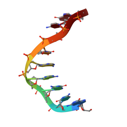
High-resolution structure of a DNA helix forming (C.G)*G base triplets.

Van Meervelt, L.Vlieghe, D.Dautant, A.Gallois, B.Precigoux, G.Kennard, O.

(1995) Nature 374: 742-744

  • DOI: https://doi.org/10.1038/374742a0
  • Primary Citation of Related Structures:  
    208D

  • PubMed Abstract: 

    Triple helices result from interaction between single- and double-stranded nucleic acids. Their formation is a possible mechanism for recombination of homologous gene sequences in nature and provides, inter alia, a basis for artificial control of gene activity. Triple-helix motifs have been extensively studied by a variety of techniques, but few high-resolution structural data are available. The only triplet structures characterized so far by X-ray diffraction were in protein-DNA complexes studied at about 3 A resolution. We report here the X-ray analysis of a DNA nonamer, d(GCGAATTCG), to a resolution of 2.05 A, in which the extended crystal structure contains (C.G)*G triplets as a fragment of triple helix. The guanosine-containing chains are in a parallel orientation. This arrangement is a necessary feature of models for homologous recombination which results ultimately in replacement of one length of DNA by another of similar sequence. The present-structure agrees with many published predictions of triplex organization, and provides an accurate representation of an element that allows sequence-specific association between single- and double-stranded nucleic acids.


  • Organizational Affiliation

    Department of Chemistry, Katholieke Universiteit Leuven, Heverlee, Belgium.


Macromolecules

Find similar nucleic acids by:  Sequence   |   3D Structure  

Entity ID: 1
MoleculeChains LengthOrganismImage
DNA (5'-D(*GP*CP*GP*AP*AP*TP*TP*CP*G)-3')
A, B
9N/A
Sequence Annotations
Expand
  • Reference Sequence
Small Molecules
Ligands 1 Unique
IDChains Name / Formula / InChI Key2D Diagram3D Interactions
MG
Query on MG

Download Ideal Coordinates CCD File 
C [auth A],
D [auth A]
MAGNESIUM ION
Mg
JLVVSXFLKOJNIY-UHFFFAOYSA-N
Experimental Data & Validation

Experimental Data

  • Method: X-RAY DIFFRACTION
  • Resolution: 2.05 Å
  • R-Value Work: 0.168 
  • R-Value Observed: 0.168 
  • Space Group: P 21 21 21
Unit Cell:
Length ( Å )Angle ( ˚ )
a = 22.238α = 90
b = 37.01β = 90
c = 54.1γ = 90
Software Package:
Software NamePurpose
AMoREphasing
X-PLORrefinement
IPMOSFLMdata reduction

Structure Validation

View Full Validation Report


Entry History 

Deposition Data

Revision History  (Full details and data files)

  • Version 1.0: 1995-09-15
    Type: Initial release
  • Version 1.1: 2008-05-22
    Changes: Version format compliance
  • Version 1.2: 2011-07-13
    Changes: Version format compliance
  • Version 1.3: 2024-02-14
    Changes: Data collection, Database references, Derived calculations
  • Version 1.4: 2024-04-03
    Changes: Refinement description