1W0N

Structure of uncomplexed Carbohydrate Binding Domain CBM36


Experimental Data Snapshot

  • Method: X-RAY DIFFRACTION
  • Resolution: 0.80 Å
  • R-Value Free: 0.144 
  • R-Value Work: 0.129 
  • R-Value Observed: 0.129 

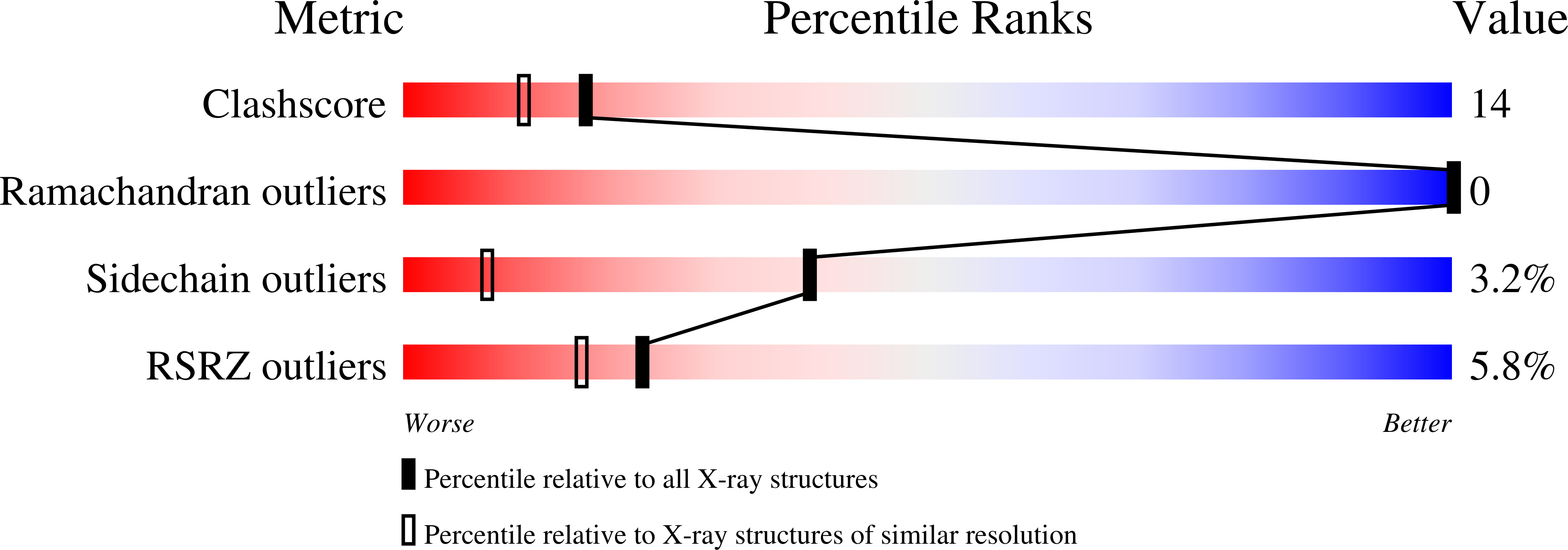
wwPDB Validation   3D Report Full Report


This is version 1.3 of the entry. See complete history


Literature

Ab Initio Structure Determination and Functional Characterization of Cbm36: A New Family of Calcium-Dependent Carbohydrate Binding Modules

Jamal, S.Boraston, A.B.Turkenburg, J.P.Tarbouriech, N.Ducros, V.M.-A.Davies, G.J.

(2004) Structure 12: 1177

  • DOI: https://doi.org/10.1016/j.str.2004.04.022
  • Primary Citation of Related Structures:  
    1UX7, 1W0N

  • PubMed Abstract: 

    The enzymatic degradation of polysaccharides harnesses multimodular enzymes whose carbohydrate binding modules (CBM) target the catalytic domain onto the recalcitrant substrate. Here we report the ab initio structure determination and subsequent refinement, at 0.8 A resolution, of the CBM36 domain of the Paenibacillus polymyxa xylanase 43A. Affinity electrophoresis, isothermal titration calorimetry, and UV difference spectroscopy demonstrate that CBM36 is a novel Ca(2+)-dependent xylan binding domain. The 3D structure of CBM36 in complex with xylotriose and Ca(2+), at 1.5 A resolution, displays significant conformational changes compared to the native structure and reveals the molecular basis for its unique Ca(2+)-dependent binding of xylooligosaccharides through coordination of the O2 and O3 hydroxyls. CBM36 is one of an emerging spectrum of carbohydrate binding modules that increasingly find applications in industry and display great potential for mapping the "glyco-architecture" of plant cells.


  • Organizational Affiliation

    Structural Biology Laboratory, Department of Chemistry, The University of York, Heslington, York YO10 5YW, United Kingdom.


Macromolecules
Find similar proteins by:  (by identity cutoff)  |  3D Structure
Entity ID: 1
MoleculeChains Sequence LengthOrganismDetailsImage
ENDO-1,4-BETA-XYLANASE D131Paenibacillus polymyxaMutation(s): 0 
EC: 3.2.1.8
UniProt
Find proteins for P45796 (Paenibacillus polymyxa)
Explore P45796 
Go to UniProtKB:  P45796
Entity Groups  
Sequence Clusters30% Identity50% Identity70% Identity90% Identity95% Identity100% Identity
UniProt GroupP45796
Sequence Annotations
Expand
  • Reference Sequence
Experimental Data & Validation

Experimental Data

  • Method: X-RAY DIFFRACTION
  • Resolution: 0.80 Å
  • R-Value Free: 0.144 
  • R-Value Work: 0.129 
  • R-Value Observed: 0.129 
  • Space Group: P 21 21 21
Unit Cell:
Length ( Å )Angle ( ˚ )
a = 30.529α = 90
b = 40.187β = 90
c = 81.2γ = 90
Software Package:
Software NamePurpose
REFMACrefinement
MOSFLMdata reduction
SCALAdata scaling

Structure Validation

View Full Validation Report


Entry History 

Deposition Data

Revision History  (Full details and data files)

  • Version 1.0: 2004-10-27
    Type: Initial release
  • Version 1.1: 2011-05-08
    Changes: Version format compliance
  • Version 1.2: 2011-07-13
    Changes: Version format compliance
  • Version 1.3: 2019-05-08
    Changes: Data collection, Experimental preparation, Other