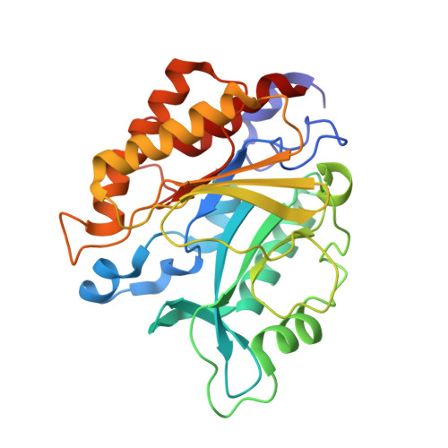
X-ray Structure of the R69D Phosphatidylinositol-Specific Phospholipase C Enzyme: Insight into the Role of Calcium and Surrounding Amino Acids in Active Site Geometry and Catalysis.
Apiyo, D., Zhao, L., Tsai, M.-D., Selby, T.L.(2005) Biochemistry 44: 9980-9989
- PubMed: 16042375
- DOI: https://doi.org/10.1021/bi047896v
- Primary Citation of Related Structures:
1T6M - PubMed Abstract:
Phosphatidylinositol-specific phospholipase Cs (PLCs) are a family of phosphodiesterases that catalyze the cleavage of the P-O bond via transesterification using the internal hydroxyl group of the substrate as a nucleophile, generating the five-membered cyclic inositol phosphate as an intermediate or product. To better understand the role of calcium in the catalytic mechanism of PLCs, we have determined the X-ray crystal structure of an engineered PLC enzyme from Bacillus thuringiensis to 2.1 A resolution. The active site of this enzyme has been altered by substituting the catalytic arginine with an aspartate at position 69 (R69D). This single-amino acid substitution converted a metal-independent, low-molecular weight enzyme into a metal ion-dependent bacterial PLC with an active site architecture similar to that of the larger metal ion-dependent mammalian PLC. The Ca(2+) ion shows a distorted square planar geometry in the active site that allows for efficient substrate binding and transition state stabilization during catalysis. Additional changes in the positions of the catalytic general acid/general base (GA/GB) were also observed, indicating the interrelation of the intricate hydrogen bonding network involved in stabilizing the active site amino acids. The functional information provided by this X-ray structure now allows for a better understanding of the catalytic mechanism, including stereochemical effects and substrate interactions, which facilitates better inhibitor design and sheds light on the possibilities of understanding how protein evolution might have occurred across this enzyme family.
Organizational Affiliation:
Department of Chemistry, The University of Central Florida, Orlando, Florida 32816, USA.