1GXS

Crystal Structure of Hydroxynitrile Lyase from Sorghum bicolor in Complex with Inhibitor Benzoic Acid: a novel cyanogenic enzyme


Experimental Data Snapshot

  • Method: X-RAY DIFFRACTION
  • Resolution: 2.30 Å
  • R-Value Free: 0.222 
  • R-Value Work: 0.165 
  • R-Value Observed: 0.165 

wwPDB Validation   3D Report Full Report


Ligand Structure Quality Assessment 


This is version 2.1 of the entry. See complete history


Literature

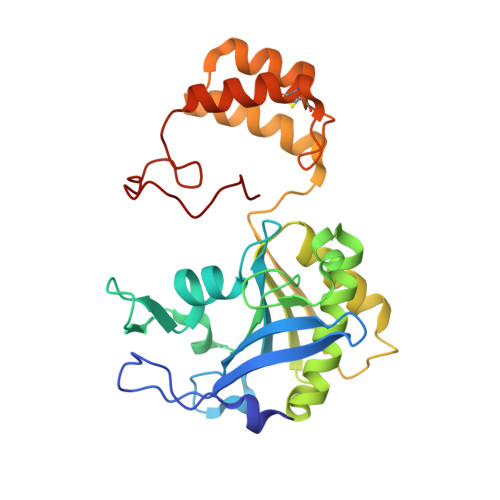
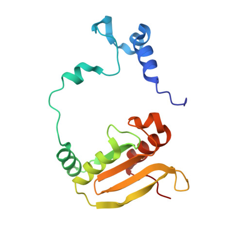
Crystal Structure of Hydroxynitrile Lyase from Sorghum Bicolor in Complex with the Inhibitor Benzoic Acid: A Novel Cyanogenic Enzyme

Lauble, H.Miehlich, B.Foerster, S.Wajant, H.Effenberger, F.

(2002) Biochemistry 41: 12043

  • DOI: https://doi.org/10.1021/bi020300o
  • Primary Citation of Related Structures:  
    1GXS

  • PubMed Abstract: 

    The crystal structure of the hydroxynitrile lyase from Sorghum bicolor (SbHNL) in complex with the inhibitor benzoic acid has been determined at 2.3 A resolution and refined to a crystallographic R-factor of 16.5%. The SbHNL sequence places the enzyme in the alpha/beta hydrolase family where the active site nucleophile is predicted to be organized in a characteristic pentapeptide motif which is part of the active site strand-turn-helix motif. In SbHNL, however, a unique two-amino acid deletion is next to the putative active site Ser158, removing thereby the putative oxyanion hole-forming Tyr residue. The presented X-ray structure shows that the overall folding pattern of SbHNL is similar to that of the closely related wheat serine carboxypeptidase (CPD-WII); however, the deletion in SbHNL is forcing the putative active site residues away from the expected hydrolase binding site toward a small hydrophobic cleft, which also contains the inhibitor benzoic acid, defining thereby a completely different SbHNL active site architecture where the traditional view of a classic triad is not given any more. Rather, we propose a mechanism involving general base catalysis by the carboxy-terminal Trp270 carboxyl group and proton transfer toward the leaving nitrile group by an active site water molecule. The unexpected interactions of the inhibitor with the new SbHNL active site also reveal the structural basis for the enzyme's limited substrate specificity. The implications of this structure on the evolution of catalysis in the hydroxynitrile lyase superfamily are discussed.


  • Organizational Affiliation

    Institut für Organische Chemie, Universität Stuttgart, Pfaffenwaldring 55, D-70569 Stuttgart, Germany. PeterLauble@t-online.de


Macromolecules
Find similar proteins by:  (by identity cutoff)  |  3D Structure
Entity ID: 1
MoleculeChains Sequence LengthOrganismDetailsImage
P-(S)-HYDROXYMANDELONITRILE LYASE CHAIN A
A, C
270Sorghum bicolorMutation(s): 4 
EC: 4.1.2.11
UniProt
Find proteins for P52708 (Sorghum bicolor)
Explore P52708 
Go to UniProtKB:  P52708
Entity Groups  
Sequence Clusters30% Identity50% Identity70% Identity90% Identity95% Identity100% Identity
UniProt GroupP52708
Sequence Annotations
Expand
  • Reference Sequence
Find similar proteins by:  (by identity cutoff)  |  3D Structure
Entity ID: 2
MoleculeChains Sequence LengthOrganismDetailsImage
P-(S)-HYDROXYMANDELONITRILE LYASE CHAIN B
B, D
158Sorghum bicolorMutation(s): 3 
EC: 4.1.2.11
UniProt
Find proteins for P52708 (Sorghum bicolor)
Explore P52708 
Go to UniProtKB:  P52708
Entity Groups  
Sequence Clusters30% Identity50% Identity70% Identity90% Identity95% Identity100% Identity
UniProt GroupP52708
Sequence Annotations
Expand
  • Reference Sequence
Oligosaccharides

Help

Entity ID: 3
MoleculeChains Length2D Diagram Glycosylation3D Interactions
beta-L-fucopyranose-(1-3)-[2-acetamido-2-deoxy-beta-D-glucopyranose-(1-4)]2-acetamido-2-deoxy-beta-D-glucopyranose
E, F
3N-Glycosylation
Glycosylation Resources
GlyTouCan:  G46874CY
GlyCosmos:  G46874CY
GlyGen:  G46874CY
Experimental Data & Validation

Experimental Data

  • Method: X-RAY DIFFRACTION
  • Resolution: 2.30 Å
  • R-Value Free: 0.222 
  • R-Value Work: 0.165 
  • R-Value Observed: 0.165 
  • Space Group: C 1 2 1
Unit Cell:
Length ( Å )Angle ( ˚ )
a = 150.7α = 90
b = 103.7β = 101.3
c = 90.6γ = 90
Software Package:
Software NamePurpose
X-PLORrefinement
MOSFLMdata reduction
SCALEPACKdata scaling
X-PLORphasing

Structure Validation

View Full Validation Report


Ligand Structure Quality Assessment 


Entry History 

Deposition Data

Revision History  (Full details and data files)

  • Version 1.0: 2002-10-01
    Type: Initial release
  • Version 1.1: 2011-10-26
    Changes: Atomic model, Database references, Derived calculations, Non-polymer description, Other, Structure summary, Version format compliance
  • Version 2.0: 2020-07-29
    Type: Remediation
    Reason: Carbohydrate remediation
    Changes: Atomic model, Data collection, Derived calculations, Other, Structure summary
  • Version 2.1: 2023-12-13
    Changes: Data collection, Database references, Refinement description, Structure summary