1QBV

CRYSTAL STRUCTURE OF THROMBIN COMPLEXED WITH AN GUANIDINE-MIMETIC INHIBITOR


Experimental Data Snapshot

  • Method: X-RAY DIFFRACTION
  • Resolution: 1.80 Å
  • R-Value Work: 0.193 
  • R-Value Observed: 0.193 

wwPDB Validation   3D Report Full Report


This is version 1.5 of the entry. See complete history


Literature

Structural analysis of thrombin complexed with potent inhibitors incorporating a phenyl group as a peptide mimetic and aminopyridines as guanidine substitutes.

Bone, R.Lu, T.Illig, C.R.Soll, R.M.Spurlino, J.C.

(1998) J Med Chem 41: 2068-2075

  • DOI: https://doi.org/10.1021/jm970796l
  • Primary Citation of Related Structures:  
    1QBV

  • PubMed Abstract: 

    The structure of the noncovalent complex of human alpha-thrombin with a nonpeptide inhibitor containing a central phenyl scaffold, N-[2-[5-methyl-3-(2-chlorophenylsulfonyloxy)phenoxy]ethyl]-N- methyl-4 -aminopyridine (1), has been determined to 2.20 A resolution. In addition, the thrombin-bound structures of two distinct amino acid-based inhibitors (3 and 4) containing different aminopyridine-derived guanidine mimetics have been determined. Each compound occupies the same region of the active site and projects an aminopyridine, a central hydrophobic group, and an aryl group, into the S1, S2, and aryl subsites on thrombin. Nonpeptide 1 forms only one direct intermolecular hydrogen bond to the thrombin active site and forms no hydrogen bonds to ordered molecules of solvent. Close contacts are observed between main-chain carbonyl groups on thrombin and the edges of the central phenyl and aminopyridine rings and the sulfonyl group of 1 such that atoms carrying opposite partial charges are juxtaposed. Aminopyridine groups in 3 and 4 also form close contacts with the edges of carbonyl groups on thrombin and are flexibly accommodated in the S1 subsite. Superposition of the bound conformations of 1 and D-Phe-Pro-amidobutylguanidine (2) revealed that the central phenyl scaffold of 1 substitutes for the peptide main chain of 2.


  • Organizational Affiliation

    3-Dimensional Pharmaceuticals Inc., Eagleview Corporate Center, 665 Stockton Drive, Suite 104, Exton, Pennsylvania 19341, USA.


Macromolecules
Find similar proteins by:  (by identity cutoff)  |  3D Structure
Entity ID: 1
MoleculeChains Sequence LengthOrganismDetailsImage
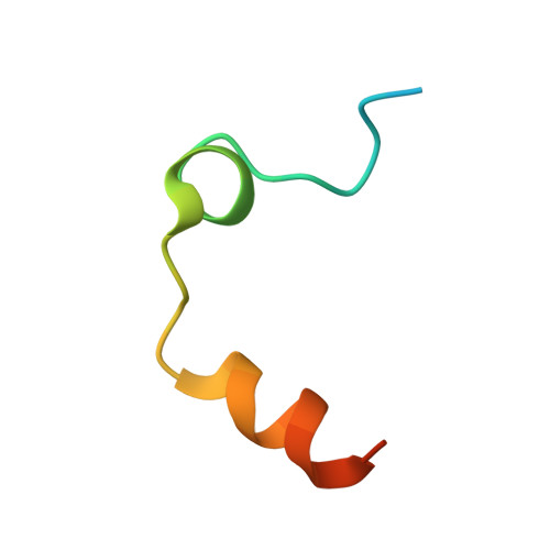
THROMBIN (LIGHT CHAIN)A [auth L]36Homo sapiensMutation(s): 0 
EC: 3.4.21.5
UniProt & NIH Common Fund Data Resources
Find proteins for P00734 (Homo sapiens)
Explore P00734 
Go to UniProtKB:  P00734
PHAROS:  P00734
GTEx:  ENSG00000180210 
Entity Groups  
Sequence Clusters30% Identity50% Identity70% Identity90% Identity95% Identity100% Identity
UniProt GroupP00734
Sequence Annotations
Expand
  • Reference Sequence
Find similar proteins by:  (by identity cutoff)  |  3D Structure
Entity ID: 2
MoleculeChains Sequence LengthOrganismDetailsImage
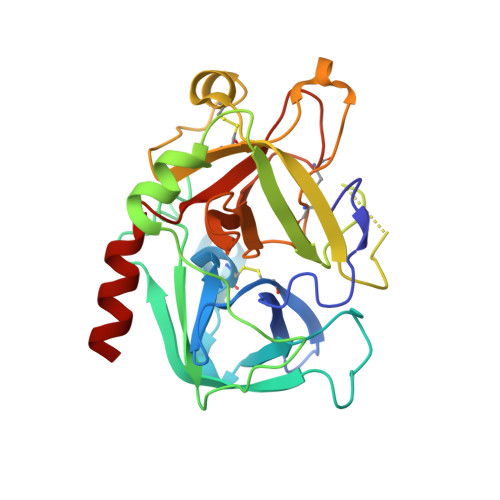
THROMBIN (HEAVY CHAIN)B [auth H]259Homo sapiensMutation(s): 0 
EC: 3.4.21.5
UniProt & NIH Common Fund Data Resources
Find proteins for P00734 (Homo sapiens)
Explore P00734 
Go to UniProtKB:  P00734
PHAROS:  P00734
GTEx:  ENSG00000180210 
Entity Groups  
Sequence Clusters30% Identity50% Identity70% Identity90% Identity95% Identity100% Identity
UniProt GroupP00734
Sequence Annotations
Expand
  • Reference Sequence

Find similar proteins by:  Sequence   |   3D Structure  

Entity ID: 3
MoleculeChains Sequence LengthOrganismDetailsImage
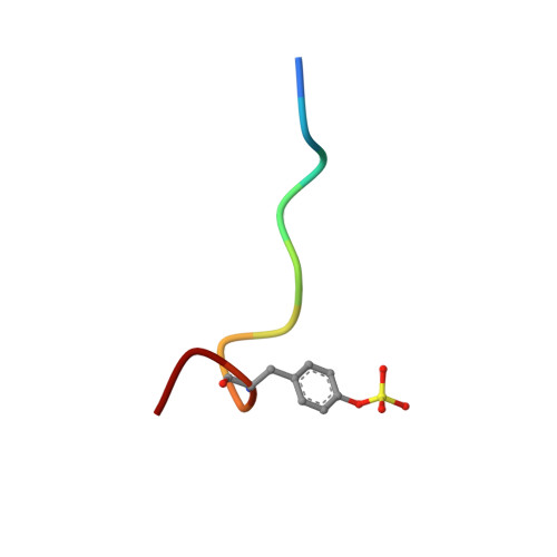
HirudinC [auth E]11Hirudo medicinalisMutation(s): 0 
UniProt
Find proteins for P28504 (Hirudo medicinalis)
Explore P28504 
Go to UniProtKB:  P28504
Entity Groups  
Sequence Clusters30% Identity50% Identity70% Identity90% Identity95% Identity100% Identity
UniProt GroupP28504
Sequence Annotations
Expand
  • Reference Sequence
Small Molecules
Ligands 1 Unique
IDChains Name / Formula / InChI Key2D Diagram3D Interactions
PPX
Query on PPX

Download Ideal Coordinates CCD File 
D [auth H][PHENYLALANINYL-PROLINYL]-[2-(PYRIDIN-4-YLAMINO)-ETHYL]-AMINE
C21 H27 N5 O2
GEIDRYBJURDUSW-MOPGFXCFSA-N
Modified Residues  1 Unique
IDChains TypeFormula2D DiagramParent
TYS
Query on TYS
C [auth E]L-PEPTIDE LINKINGC9 H11 N O6 STYR
Binding Affinity Annotations 
IDSourceBinding Affinity
PPX Binding MOAD:  1QBV Ki: 4100 (nM) from 1 assay(s)
PDBBind:  1QBV Ki: 4100 (nM) from 1 assay(s)
BindingDB:  1QBV Ki: 4100 (nM) from 1 assay(s)
Experimental Data & Validation

Experimental Data

  • Method: X-RAY DIFFRACTION
  • Resolution: 1.80 Å
  • R-Value Work: 0.193 
  • R-Value Observed: 0.193 
  • Space Group: C 1 2 1
Unit Cell:
Length ( Å )Angle ( ˚ )
a = 70.9α = 90
b = 71.8β = 100.52
c = 72.7γ = 90
Software Package:
Software NamePurpose
X-PLORmodel building
X-PLORrefinement
X-PLORphasing

Structure Validation

View Full Validation Report


Entry History 

Deposition Data

Revision History  (Full details and data files)

  • Version 1.0: 1999-10-27
    Type: Initial release
  • Version 1.1: 2008-04-27
    Changes: Version format compliance
  • Version 1.2: 2011-07-13
    Changes: Atomic model, Database references, Derived calculations, Non-polymer description, Structure summary, Version format compliance
  • Version 1.3: 2012-12-12
    Changes: Other
  • Version 1.4: 2013-03-13
    Changes: Other
  • Version 1.5: 2017-10-04
    Changes: Advisory, Refinement description