7USM | pdb_00007usm

Integrin alphaM/beta2 ectodomain


Experimental Data Snapshot

  • Method: ELECTRON MICROSCOPY
  • Resolution: 2.70 Å
  • Aggregation State: PARTICLE 
  • Reconstruction Method: SINGLE PARTICLE 

wwPDB Validation 3D Report Full Report

Validation slider image for 7USM

This is version 1.2 of the entry. See complete history

Literature

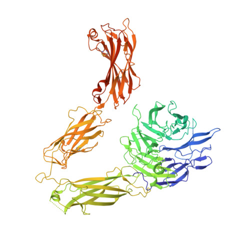
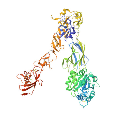
Structural basis for non-canonical integrin engagement by Bordetella adenylate cyclase toxin.

Goldsmith, J.A.DiVenere, A.M.Maynard, J.A.McLellan, J.S.

(2022) Cell Rep 40: 111196-111196

  • DOI: https://doi.org/10.1016/j.celrep.2022.111196
  • Primary Citation Related Structures: 
    7USL, 7USM

  • PubMed Abstract: 

    Integrins are ubiquitous cell-surface heterodimers that are exploited by pathogens and toxins, including leukotoxins that target β 2 integrins on phagocytes. The Bordetella adenylate cyclase toxin (ACT) uses the α M β 2 integrin as a receptor, but the structural basis for integrin binding and neutralization by antibodies is poorly understood. Here, we use cryoelectron microscopy to determine a 2.7 Å resolution structure of an ACT fragment bound to α M β 2 . This structure reveals that ACT interacts with the headpiece and calf-2 of the α M subunit in a non-canonical manner specific to bent, inactive α M β 2 . Neutralizing antibody epitopes map to ACT residues involved in α M binding, providing the basis for antibody-mediated attachment inhibition. Furthermore, binding to α M β 2 positions the essential ACT acylation sites, which are conserved among toxins exported by type I secretion systems, at the cell membrane. These findings reveal a structural mechanism for integrin-mediated attachment and explain antibody-mediated neutralization of ACT intoxication.


  • Organizational Affiliation
    • Department of Molecular Biosciences, The University of Texas at Austin, Austin, TX 78712, USA.

Macromolecule Content 

  • Total Structure Weight: 211.82 kDa 
  • Atom Count: 11,893 
  • Modeled Residue Count: 1,521 
  • Deposited Residue Count: 1,892 
  • Unique protein chains: 2

Macromolecules

Find similar proteins by:|  3D Structure
Entity ID: 1
MoleculeChains  Sequence LengthOrganismDetailsImage
Integrin alpha-M1,162Homo sapiensMutation(s): 0 
Gene Names: ITGAMCD11BCR3A
UniProt & NIH Common Fund Data Resources
Find proteins for P11215 (Homo sapiens)
Explore P11215 
Go to UniProtKB:  P11215
PHAROS:  P11215
GTEx:  ENSG00000169896 
Entity Groups
Sequence Clusters30% Identity50% Identity70% Identity90% Identity95% Identity100% Identity
UniProt GroupP11215
Glycosylation
Glycosylation Sites: 9Go to GlyGen: P11215-1
Sequence Annotations
Expand
Reference Sequence
Find similar proteins by:|  3D Structure
Entity ID: 2
MoleculeChains  Sequence LengthOrganismDetailsImage
Integrin beta730Homo sapiensMutation(s): 0 
Gene Names: ITGB2
UniProt & NIH Common Fund Data Resources
Find proteins for P05107 (Homo sapiens)
Explore P05107 
Go to UniProtKB:  P05107
PHAROS:  P05107
GTEx:  ENSG00000160255 
Entity Groups
Sequence Clusters30% Identity50% Identity70% Identity90% Identity95% Identity100% Identity
UniProt GroupP05107
Glycosylation
Glycosylation Sites: 1Go to GlyGen: P05107-1
Sequence Annotations
Expand
Reference Sequence

Oligosaccharides

Help  
Entity ID: 3
MoleculeChains Length2D Diagram GlycosylationD Interactions
2-acetamido-2-deoxy-beta-D-glucopyranose-(1-4)-2-acetamido-2-deoxy-beta-D-glucopyranose
C
2N-Glycosylation
Glycosylation Resources
GlyTouCan: G42666HT
GlyCosmos: G42666HT
GlyGen: G42666HT

Small Molecules

Ligands 2 Unique
IDChains Name / Formula / InChI Key2D Diagram3D Interactions
NAG

Query on NAG


Download:Ideal Coordinates CCD File
G [auth A]
H [auth A]
I [auth A]
J [auth A]
K [auth A]
G [auth A],
H [auth A],
I [auth A],
J [auth A],
K [auth A],
L [auth A],
M [auth A],
N [auth A],
P [auth B]
2-acetamido-2-deoxy-beta-D-glucopyranose
C8 H15 N O6
OVRNDRQMDRJTHS-FMDGEEDCSA-N
CA

Query on CA


Download:Ideal Coordinates CCD File
D [auth A],
E [auth A],
F [auth A],
O [auth B]
CALCIUM ION
Ca
BHPQYMZQTOCNFJ-UHFFFAOYSA-N

Experimental Data & Validation

Experimental Data

  • Method: ELECTRON MICROSCOPY
  • Resolution: 2.70 Å
  • Aggregation State: PARTICLE 
  • Reconstruction Method: SINGLE PARTICLE 

Structure Validation

View Full Validation Report


Entry History 

& Funding Information

Deposition Data


Funding OrganizationLocationGrant Number
National Institutes of Health/National Institute Of Allergy and Infectious Diseases (NIH/NIAID)United StatesAI155453

Revision History  (Full details and data files)

  • Version 1.0: 2022-08-17
    Type: Initial release
  • Version 1.1: 2022-09-14
    Changes: Database references
  • Version 1.2: 2024-10-30
    Changes: Data collection, Structure summary