9M5V | pdb_00009m5v

Structure of human TRPC5 bound with (-)-englerin A,class1


Experimental Data Snapshot

  • Method: ELECTRON MICROSCOPY
  • Resolution: 2.53 Å
  • Aggregation State: PARTICLE 
  • Reconstruction Method: SINGLE PARTICLE 

wwPDB Validation   3D Report Full Report


This is version 1.1 of the entry. See complete history


Literature

Mechanism of (-)-Englerin A and calcium binding on the human TRPC5 channel.

Chen, Y.Song, K.Guo, W.Wei, M.Chen, L.

(2025) Protein Sci 34: e70218-e70218

  • DOI: https://doi.org/10.1002/pro.70218
  • Primary Citation of Related Structures:  
    9M36, 9M4W, 9M5V

  • PubMed Abstract: 

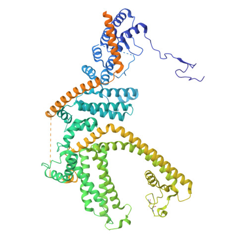
    The natural product (-)-Englerin A (EA) selectively inhibits renal cancer cell growth by potently activating TRPC4 and TRPC5-containing ion channels. However, its binding site on these channels has remained elusive. In this study, we present two cryo-EM structures of human TRPC5 in complex with EA at 2.5 and 2.6 Å resolution, which reveal the EA-binding site and identify two major conformations influenced by calcium. EA binds between the pore helix and S5/S6 helices of hTRPC5, forming critical hydrophobic and polar interactions that underscore its specificity. Calcium binding at the intracellular domain of TRPC5 induces structural changes that stabilize the domain in a compact conformation. These findings expand our understanding of the structural pharmacology of TRPC5 and provide a framework for investigating calcium regulation in TRPC channels.


  • Organizational Affiliation
    • State Key Laboratory of Membrane Biology, College of Future Technology, Institute of Molecular Medicine, Beijing Key Laboratory of Cardiometabolic Molecular Medicine, Peking University, Beijing, China.

Macromolecules
Find similar proteins by:  (by identity cutoff)  |  3D Structure
Entity ID: 1
MoleculeChains Sequence LengthOrganismDetailsImage
Short transient receptor potential channel 5
A, B, C, D
976Homo sapiensMutation(s): 0 
Gene Names: TRPC5TRP5
UniProt & NIH Common Fund Data Resources
Find proteins for Q9UL62 (Homo sapiens)
Explore Q9UL62 
Go to UniProtKB:  Q9UL62
PHAROS:  Q9UL62
GTEx:  ENSG00000072315 
Entity Groups  
Sequence Clusters30% Identity50% Identity70% Identity90% Identity95% Identity100% Identity
UniProt GroupQ9UL62
Sequence Annotations
Expand
  • Reference Sequence
Small Molecules
Ligands 5 Unique
IDChains Name / Formula / InChI Key2D Diagram3D Interactions
PTY
Query on PTY

Download Ideal Coordinates CCD File 
H [auth A],
M [auth B],
R [auth C],
W [auth D]
PHOSPHATIDYLETHANOLAMINE
C40 H80 N O8 P
NJGIRBISCGPRPF-KXQOOQHDSA-N
Y01
Query on Y01

Download Ideal Coordinates CCD File 
E [auth A],
J [auth B],
O [auth C],
T [auth D]
CHOLESTEROL HEMISUCCINATE
C31 H50 O4
WLNARFZDISHUGS-MIXBDBMTSA-N
A1L55
Query on A1L55

Download Ideal Coordinates CCD File 
I [auth A],
N [auth B],
S [auth C],
X [auth D]
(-)-englerin A
C26 H34 O6
GACOFEKSDCOVMV-RRYXBOBMSA-N
ZN
Query on ZN

Download Ideal Coordinates CCD File 
F [auth A],
K [auth B],
P [auth C],
U [auth D]
ZINC ION
Zn
PTFCDOFLOPIGGS-UHFFFAOYSA-N
CA
Query on CA

Download Ideal Coordinates CCD File 
G [auth A],
L [auth B],
Q [auth C],
V [auth D]
CALCIUM ION
Ca
BHPQYMZQTOCNFJ-UHFFFAOYSA-N
Experimental Data & Validation

Experimental Data

  • Method: ELECTRON MICROSCOPY
  • Resolution: 2.53 Å
  • Aggregation State: PARTICLE 
  • Reconstruction Method: SINGLE PARTICLE 

Structure Validation

View Full Validation Report


Entry History & Funding Information

Deposition Data


Funding OrganizationLocationGrant Number
Ministry of Science and Technology (MoST, China)China2022YFA0806504
National Natural Science Foundation of China (NSFC)China32225027

Revision History  (Full details and data files)

  • Version 1.0: 2025-07-02
    Type: Initial release
  • Version 1.1: 2025-11-12
    Changes: Data collection, Database references