9LRC | pdb_00009lrc

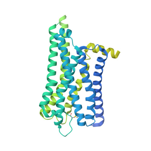
Cryo-EM structure of the histamine H4 receptor-Gi protein complex (Receptor focused)


Experimental Data Snapshot

  • Method: ELECTRON MICROSCOPY
  • Resolution: 2.84 Å
  • Aggregation State: PARTICLE 
  • Reconstruction Method: SINGLE PARTICLE 

wwPDB Validation 3D Report Full Report

Validation slider image for 9LRC

This is version 1.1 of the entry. See complete history. 

Literature

Structural insights into ligand recognition and G protein preferences across histamine receptors.

Matsuzaki, Y., Sano, F.K., Oshima, H.S., Akasaka, H., Kobayashi, K., Tanaka, T., Itoh, Y., Shihoya, W., Kise, Y., Kusakizako, T., Inoue, A., Nureki, O.

(2025) Commun Biol 8: 957-957

  • DOI: https://doi.org/10.1038/s42003-025-08363-7
  • Primary Citation Related Structures: 
    9LRB, 9LRC, 9LRD, 9LRE

  • PubMed Abstract: 

    Histamine exerts critical physiological roles by activating four receptor subtypes, each exhibiting a specific G protein preference. Among these, the histamine H 4 receptor (H 4 R) modulates chemotaxis and interferon production through G i protein activation, suggesting its therapeutic potential. Despite its physiological significance, the mechanisms underlying H 4 R signalling and G protein preference across histamine receptors remain poorly understood. Here, we present the cryo-electron microscopy structure of the H 4 R-G i complex, revealing unique mechanisms of histamine recognition and receptor activation. We further solved the structures of the histamine H 1 receptor (H 1 R) bound to the non-canonical G proteins G i and G s . Through a combination of functional and computational analyses, we identified the intracellular loop 2 as a critical determinant of G protein preference in H 1 R and H 4 R. Collectively, our comprehensive study revealed the structural basis for distinct mechanisms of ligand recognition and receptor activation, offering a profound insight into G protein preference across receptor subtypes.


  • Organizational Affiliation: 
    • Department of Biological Sciences, Graduate School of Science, The University of Tokyo, Bunkyo-Ku, Tokyo, Japan.

Macromolecule Content 

  • Total Structure Weight: 77.91 kDa 
  • Atom Count: 2,328 
  • Modeled Residue Count: 282 
  • Deposited Residue Count: 692 
  • Unique protein chains: 1

Macromolecules

Find similar proteins by:|  3D Structure
Entity ID: 1
MoleculeChains  Sequence LengthOrganismDetailsImage
Histamine H4 receptor,Genome polyproteinA [auth R]692Homo sapiensMutation(s): 0 
Gene Names: HRH4, GPCR105
EC: 3.4.22.29 (PDB Primary Data), 3.6.1.15 (PDB Primary Data), 3.4.22.28 (PDB Primary Data), 2.7.7.48 (PDB Primary Data)
Membrane Entity: Yes 
UniProt & NIH Common Fund Data Resources
Find proteins for Q9H3N8 (Homo sapiens)
Explore Q9H3N8 
Go to UniProtKB:  Q9H3N8
PHAROS:  Q9H3N8
GTEx:  ENSG00000134489 
Entity Groups
Sequence Clusters30% Identity50% Identity70% Identity90% Identity95% Identity100% Identity
UniProt GroupQ9H3N8
Sequence Annotations
Expand
Reference Sequence

Small Molecules

Ligands 1 Unique
IDChains Name / Formula / InChI Key2D Diagram3D Interactions
HSM
(Subject of Investigation/LOI)

Query on HSM


Download:Ideal Coordinates CCD File
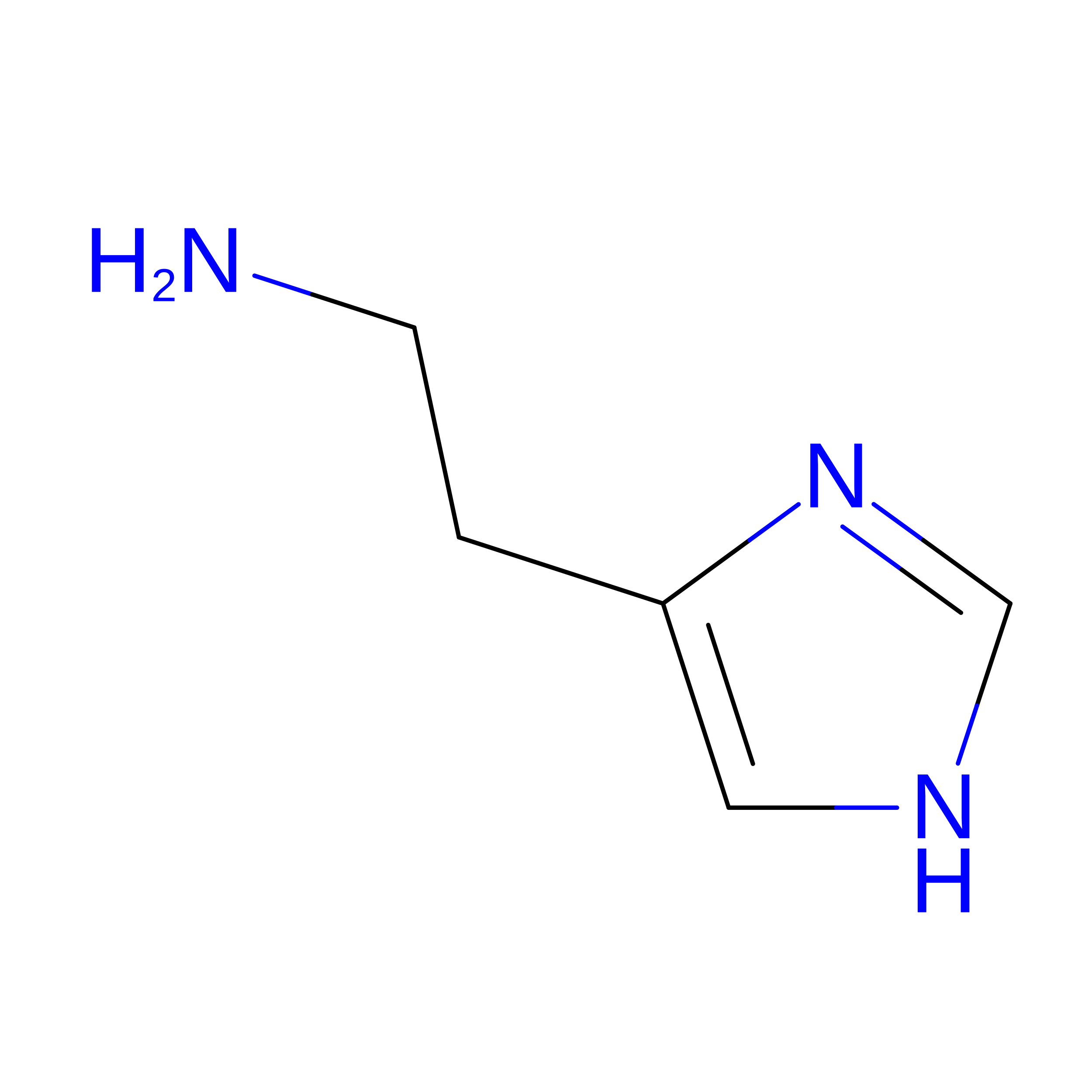
B [auth R]HISTAMINE
C5 H9 N3
NTYJJOPFIAHURM-UHFFFAOYSA-N

Experimental Data & Validation

Experimental Data

  • Method: ELECTRON MICROSCOPY
  • Resolution: 2.84 Å
  • Aggregation State: PARTICLE 
  • Reconstruction Method: SINGLE PARTICLE 
EM Software:
TaskSoftware PackageVersion
RECONSTRUCTIONcryoSPARC

Structure Validation

View Full Validation Report


Entry History 

& Funding Information

Deposition Data


Funding OrganizationLocationGrant Number
Japan Society for the Promotion of Science (JSPS)Japan--

Revision History  (Full details and data files)

  • Version 1.0: 2025-06-11
    Type: Initial release
  • Version 1.1: 2025-07-23
    Changes: Data collection, Database references