9HLS | pdb_00009hls

Structure of A16/G9 (vaccinia virus) in complex with VHH D07, VHH B01 and VHH C05


Experimental Data Snapshot

  • Method: X-RAY DIFFRACTION
  • Resolution: 3.20 Å
  • R-Value Free: 
    0.292 (Depositor), 0.293 (DCC) 
  • R-Value Work: 
    0.237 (Depositor), 0.237 (DCC) 
  • R-Value Observed: 
    0.239 (Depositor) 

Starting Models: experimental, in silico
View more details

wwPDB Validation   3D Report Full Report


Ligand Structure Quality Assessment 


This is version 1.2 of the entry. See complete history


Literature

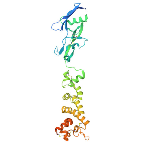
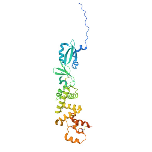
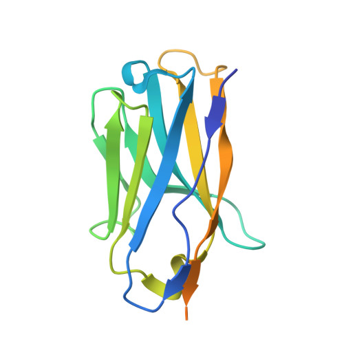
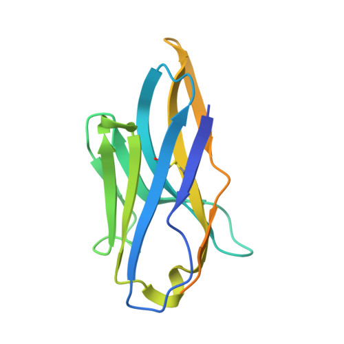
Structural basis of poxvirus fusion regulation and anti-A16/G9 antibody-mediated neutralization and protection.

Meola, A.Vernuccio, R.Battini, L.Albericio, G.Delgado, P.Bamford, R.Pokorny, L.Broutin, M.Martinez Leon, A.Gallien, S.Gil, M.Noriega, M.A.Guivel-Benhassine, F.Porrot, F.Postal, J.Buchrieser, J.Hubert, M.Haouz, A.Lafaye, P.Esteban, M.Hub, J.S.Mahevas, M.Chappert, P.Mercer, J.Garcia-Arriaza, J.Schwartz, O.Guardado-Calvo, P.

(2025) Cell 188: 6266

  • DOI: https://doi.org/10.1016/j.cell.2025.07.040
  • Primary Citation of Related Structures:  
    9HBK, 9HL2, 9HLS, 9HNG, 9HPA, 9R09, 9R0B, 9R0J, 9RDH

  • PubMed Abstract: 

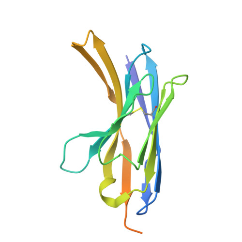
    Monkeypox virus (MPXV) is a poxvirus endemic to Central and West Africa with high epidemic potential. Poxviruses enter host cells via a conserved entry-fusion complex (EFC), which mediates viral fusion to the cell membrane. The EFC is a promising therapeutic target, but the absence of structural data has limited the development of fusion-inhibiting treatments. Here, we investigated A16/G9, a subcomplex of the EFC that controls fusion timing. Using cryo-electron microscopy, we showed how A16/G9 interacts with A56/K2, a viral fusion suppressor that prevents superinfection. Immunization with A16/G9 elicited a protective immune response in mice. Using X-ray crystallography, we characterized two neutralizing antibodies and engineered a chimeric antibody that cross-neutralizes several poxviruses more efficiently than 7D11, the most potent antibody targeting the EFC described to date. These findings highlight the potential of A16/G9 as a candidate for subunit vaccines and identify regions of the EFC as targets for antiviral development.


  • Organizational Affiliation
    • Structural biology of infectious diseases G5+ unit, Institut Pasteur, Université Paris Cité, Paris, France.

Macromolecules
Find similar proteins by:  (by identity cutoff)  |  3D Structure
Entity ID: 1
MoleculeChains Sequence LengthOrganismDetailsImage
Virion membrane protein OPG143
A, F
358Vaccinia virus Western ReserveMutation(s): 0 
Gene Names: OPG143VACWR136A16L
UniProt
Find proteins for P16710 (Vaccinia virus (strain Western Reserve))
Explore P16710 
Go to UniProtKB:  P16710
Entity Groups  
Sequence Clusters30% Identity50% Identity70% Identity90% Identity95% Identity100% Identity
UniProt GroupP16710
Sequence Annotations
Expand
  • Reference Sequence
Find similar proteins by:  (by identity cutoff)  |  3D Structure
Entity ID: 2
MoleculeChains Sequence LengthOrganismDetailsImage
Entry-fusion complex protein OPG094
B, G
334Vaccinia virus Western ReserveMutation(s): 0 
Gene Names: OPG094VACWR087G9R
UniProt
Find proteins for P07611 (Vaccinia virus (strain Western Reserve))
Explore P07611 
Go to UniProtKB:  P07611
Entity Groups  
Sequence Clusters30% Identity50% Identity70% Identity90% Identity95% Identity100% Identity
UniProt GroupP07611
Sequence Annotations
Expand
  • Reference Sequence
Find similar proteins by:  (by identity cutoff)  |  3D Structure
Entity ID: 3
MoleculeChains Sequence LengthOrganismDetailsImage
VHH D07
C, H
154Vicugna pacosMutation(s): 0 
Entity Groups  
Sequence Clusters30% Identity50% Identity70% Identity90% Identity95% Identity100% Identity
Sequence Annotations
Expand
  • Reference Sequence
Find similar proteins by:  (by identity cutoff)  |  3D Structure
Entity ID: 4
MoleculeChains Sequence LengthOrganismDetailsImage
VHH B01
D, I
156Vicugna pacosMutation(s): 0 
Entity Groups  
Sequence Clusters30% Identity50% Identity70% Identity90% Identity95% Identity100% Identity
Sequence Annotations
Expand
  • Reference Sequence
Find similar proteins by:  (by identity cutoff)  |  3D Structure
Entity ID: 5
MoleculeChains Sequence LengthOrganismDetailsImage
VHH C05E [auth J],
J [auth E]
156Vicugna pacosMutation(s): 0 
Entity Groups  
Sequence Clusters30% Identity50% Identity70% Identity90% Identity95% Identity100% Identity
Sequence Annotations
Expand
  • Reference Sequence
Experimental Data & Validation

Experimental Data

  • Method: X-RAY DIFFRACTION
  • Resolution: 3.20 Å
  • R-Value Free:  0.292 (Depositor), 0.293 (DCC) 
  • R-Value Work:  0.237 (Depositor), 0.237 (DCC) 
  • R-Value Observed: 0.239 (Depositor) 
Space Group: C 1 2 1
Unit Cell:
Length ( Å )Angle ( ˚ )
a = 150.435α = 90
b = 194.282β = 91.263
c = 94.866γ = 90
Software Package:
Software NamePurpose
PHENIXrefinement
XDSdata reduction
Aimlessdata scaling
PHASERphasing

Structure Validation

View Full Validation Report


Ligand Structure Quality Assessment 


Entry History & Funding Information

Deposition Data


Funding OrganizationLocationGrant Number
Agence Nationale de la Recherche (ANR)FranceANR-22-CE11-0003

Revision History  (Full details and data files)

  • Version 1.0: 2025-09-03
    Type: Initial release
  • Version 1.1: 2025-09-10
    Changes: Database references
  • Version 1.2: 2025-11-12
    Changes: Database references