9Y6M | pdb_00009y6m

Avermitilol synthase: Complex with Mg, inorganic pyrophosphate, and adamantane cation


Experimental Data Snapshot

  • Method: X-RAY DIFFRACTION
  • Resolution: 1.42 Å
  • R-Value Free: 
    0.201 (Depositor), 0.200 (DCC) 
  • R-Value Work: 
    0.160 (Depositor), 0.160 (DCC) 
  • R-Value Observed: 
    0.161 (Depositor) 

Starting Model: experimental
View more details

wwPDB Validation 3D Report Full Report

Validation slider image for 9Y6M

Ligand Structure Quality Assessment 


This is version 1.1 of the entry. See complete history

Literature

Structure and Mechanism of Avermitilol Synthase, a Sesquiterpene Cyclase That Generates a Highly Strained 6-6-3 Tricyclic Alcohol.

Gaynes, M.N.Osika, K.R.Christianson, D.W.

(2025) Biochemistry 64: 4830-4840

  • DOI: https://doi.org/10.1021/acs.biochem.5c00565
  • Primary Citation Related Structures: 
    9Y6I, 9Y6J, 9Y6K, 9Y6L, 9Y6M

  • PubMed Abstract: 

    Avermitilol synthase from Streptomyces avermitilis (SaAS) is a high-fidelity class I terpene cyclase that converts farnesyl diphosphate into a highly strained, 6-6-3 tricyclic sesquiterpene alcohol. The mechanism of avermitilol formation proceeds through a 10-3 bicyclic intermediate, bicyclogermacrene, which undergoes proton-initiated anti-Markovnikov addition to two separate C═C bonds in a transannulation mechanism that forms the 6-6-3 tricyclic skeleton, with quenching by water to yield avermitilol. Small amounts of a side product, viridifloral, result from Markovnikov addition to one of the reactive C═C bonds. Here, we present enzymological studies of SaAS to establish the substrate scope and metal ion dependence for catalysis, and we present crystal structures of SaAS complexed with a variety of ligands that partially mimic carbocation intermediates in catalysis. Interestingly, these structures show that two water molecules remain trapped in a polar crevice in the active site regardless of the ligand bound. Structure-activity relationships for site-specific mutants yield key insight into the catalytic importance of these trapped water molecules. Specifically, T215 normally hydrogen bonds with water molecule W1, but the T215V substitution breaks this hydrogen bond and causes W1 to shift by 1.3 Å to form a hydrogen bond with W300. Avermitilol generation is completely lost in this mutant, but the generation of viridifloral and another side product is enhanced. We conclude that the T215V substitution causes water molecule W1 to align for reaction with the tertiary and not the secondary carbon in the reactive C═C bond of bicyclogermacrene.


  • Organizational Affiliation
    • Roy and Diana Vagelos Laboratories, Department of Chemistry, University of Pennsylvania, Philadelphia, Pennsylvania 19104-6323, United States.

Macromolecule Content 

  • Total Structure Weight: 37.4 kDa 
  • Atom Count: 2,750 
  • Modeled Residue Count: 335 
  • Deposited Residue Count: 338 
  • Unique protein chains: 1

Macromolecules

Find similar proteins by:|  3D Structure
Entity ID: 1
MoleculeChains  Sequence LengthOrganismDetailsImage
Avermitilol synthase338Streptomyces avermitilisMutation(s): 0 
Gene Names: tpc1SAV_76
EC: 4.2.3.96
UniProt
Find proteins for Q82RR7 (Streptomyces avermitilis (strain ATCC 31267 / DSM 46492 / JCM 5070 / NBRC 14893 / NCIMB 12804 / NRRL 8165 / MA-4680))
Explore Q82RR7 
Go to UniProtKB:  Q82RR7
Entity Groups
Sequence Clusters30% Identity50% Identity70% Identity90% Identity95% Identity100% Identity
UniProt GroupQ82RR7
Sequence Annotations
Expand
Reference Sequence

Small Molecules

Ligands 4 Unique
IDChains Name / Formula / InChI Key2D Diagram3D Interactions
A1CSW
(Subject of Investigation/LOI)

Query on A1CSW


Download:Ideal Coordinates CCD File
C [auth A]1-(trimethyl-$l^{4}-azanyl)adamantane
C13 H24 N
KKXBPUAYFJQMLN-WNGYAQOMSA-N
DPO

Query on DPO


Download:Ideal Coordinates CCD File
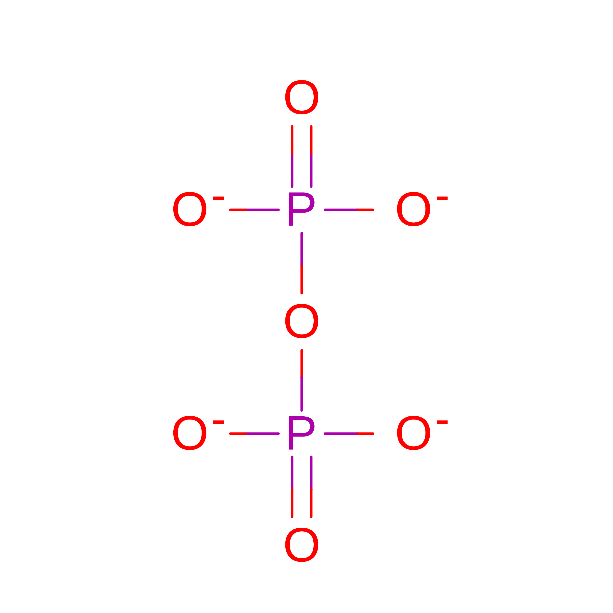
B [auth A]DIPHOSPHATE
O7 P2
XPPKVPWEQAFLFU-UHFFFAOYSA-J
EDO

Query on EDO


Download:Ideal Coordinates CCD File
D [auth A],
E [auth A]
1,2-ETHANEDIOL
C2 H6 O2
LYCAIKOWRPUZTN-UHFFFAOYSA-N
MG

Query on MG


Download:Ideal Coordinates CCD File
F [auth A],
G [auth A],
H [auth A]
MAGNESIUM ION
Mg
JLVVSXFLKOJNIY-UHFFFAOYSA-N

Experimental Data & Validation

Experimental Data

  • Method: X-RAY DIFFRACTION
  • Resolution: 1.42 Å
  • R-Value Free:  0.201 (Depositor), 0.200 (DCC) 
  • R-Value Work:  0.160 (Depositor), 0.160 (DCC) 
  • R-Value Observed: 0.161 (Depositor) 
Space Group: C 1 2 1
Unit Cell:
Length ( Å )Angle ( ˚ )
a = 95.552α = 90
b = 56.25β = 125.3
c = 72.766γ = 90
Software Package:
Software NamePurpose
PHENIXrefinement
XDSdata reduction
Aimlessdata scaling
PHASERphasing

Structure Validation

View Full Validation Report


Ligand Structure Quality Assessment 


Entry History 

& Funding Information

Deposition Data


Funding OrganizationLocationGrant Number
National Institutes of Health/National Institute of General Medical Sciences (NIH/NIGMS)United StatesGM56838

Revision History  (Full details and data files)

  • Version 1.0: 2025-12-17
    Type: Initial release
  • Version 1.1: 2025-12-31
    Changes: Database references