9Y5Q | pdb_00009y5q

Cryo EM structure of KCa3.1_R355K_I/calmodulin channel in complex with rimtuzalcap

  • Classification: TRANSPORT PROTEIN
  • Organism(s): Homo sapiens
  • Expression System: Homo sapiens
  • Mutation(s): Yes 

  • Deposited: 2025-09-05 Released: 2025-10-15 
  • Deposition Author(s): Nam, Y.W., Zhang, M.
  • Funding Organization(s): American Heart Association, National Institutes of Health/National Institute of Neurological Disorders and Stroke (NIH/NINDS)

Experimental Data Snapshot

  • Method: ELECTRON MICROSCOPY
  • Resolution: 4.73 Å
  • Aggregation State: PARTICLE 
  • Reconstruction Method: SINGLE PARTICLE 

wwPDB Validation   3D Report Full Report


This is version 1.2 of the entry. See complete history


Literature

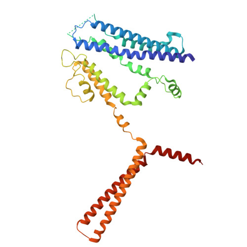
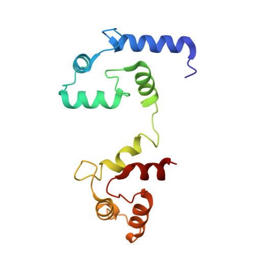
Structural basis for the subtype-selectivity of K Ca 2.2 channel activators.

Nam, Y.W.Ramanishka, A.Xu, Y.Yasuda, R.M.H.Nasburg, J.A.Im, D.Cui, M.Chandy, K.G.Wulff, H.Zhang, M.

(2026) Nat Commun 17: 531-531

  • DOI: https://doi.org/10.1038/s41467-025-67232-3
  • Primary Citation of Related Structures:  
    9O85, 9O93, 9OA8, 9Y5Q, 9YDZ

  • PubMed Abstract: 

    Small-conductance (K Ca 2.2) and intermediate-conductance (K Ca 3.1) Ca 2+ -activated K + channels are gated by a Ca 2+ -calmodulin dependent mechanism. NS309 potentiates the activity of both K Ca 2.2 and K Ca 3.1, while rimtuzalcap selectively activates K Ca 2.2. Rimtuzalcap has been used in clinical trials for the treatment of spinocerebellar ataxia and essential tremor. We report cryo-electron microscopy structures of NS309-bound K Ca 2.2 and K Ca 3.1, in addition to structures of rimtuzalcap-bound K Ca 2.2 and mutant K Ca 3.1_R355K. The different conformations of calmodulin and the cytoplasmic HC helices in the two channels underlie the subtype-selectivity of rimtuzalcap for K Ca 2.2. NS309 binds to pre-existing pockets in both channels, while the bulkier rimtuzalcap binds in an induced-fit pocket in K Ca 2.2 requiring conformational changes. In K Ca 2.2, calmodulin's N-lobes are sufficiently far apart to enable conformational changes to accommodate either NS309 or rimtuzalcap. In K Ca 3.1, calmodulin's N-lobes are closer to each other and constrained by K Ca 3.1's HC helices, which allows binding of NS309 but not rimtuzalcap. Replacement of arginine-355 in K Ca 3.1's HB helix with lysine (K Ca 3.1_R355K) allows the binding of rimtuzalcap and renders the mutant channel sensitive to rimtuzalcap. These structures provide a framework for structure-based drug design targeting K Ca 2.2 channels.


  • Organizational Affiliation
    • Department of Biomedical and Pharmaceutical Sciences, Chapman University School of Pharmacy, Irvine, CA, USA.

Macromolecules
Find similar proteins by:  (by identity cutoff)  |  3D Structure
Entity ID: 1
MoleculeChains Sequence LengthOrganismDetailsImage
Intermediate conductance calcium-activated potassium channel protein 4
A, B, C, D
378Homo sapiensMutation(s): 1 
Gene Names: KCNN4IK1IKCA1KCA4SK4
UniProt & NIH Common Fund Data Resources
Find proteins for O15554 (Homo sapiens)
Explore O15554 
Go to UniProtKB:  O15554
PHAROS:  O15554
GTEx:  ENSG00000104783 
Entity Groups  
Sequence Clusters30% Identity50% Identity70% Identity90% Identity95% Identity100% Identity
UniProt GroupO15554
Sequence Annotations
Expand
  • Reference Sequence
Find similar proteins by:  (by identity cutoff)  |  3D Structure
Entity ID: 2
MoleculeChains Sequence LengthOrganismDetailsImage
Calmodulin-1
E, F, G, H
146Homo sapiensMutation(s): 0 
Gene Names: CALM1CALMCAMCAM1
UniProt & NIH Common Fund Data Resources
Find proteins for P0DP23 (Homo sapiens)
Explore P0DP23 
Go to UniProtKB:  P0DP23
PHAROS:  P0DP23
GTEx:  ENSG00000198668 
Entity Groups  
Sequence Clusters30% Identity50% Identity70% Identity90% Identity95% Identity100% Identity
UniProt GroupP0DP23
Sequence Annotations
Expand
  • Reference Sequence
Small Molecules
Ligands 3 Unique
IDChains Name / Formula / InChI Key2D Diagram3D Interactions
A1B92 (Subject of Investigation/LOI)
Query on A1B92

Download Ideal Coordinates CCD File 
J [auth E],
L [auth F],
N [auth G],
P [auth H]
Rimtuzalcap
C18 H24 F2 N6 O
OVLIDRAJVMUEMC-UHFFFAOYSA-N
CA (Subject of Investigation/LOI)
Query on CA

Download Ideal Coordinates CCD File 
K [auth E],
M [auth F],
O [auth G],
Q [auth H]
CALCIUM ION
Ca
BHPQYMZQTOCNFJ-UHFFFAOYSA-N
K (Subject of Investigation/LOI)
Query on K

Download Ideal Coordinates CCD File 
I [auth C]POTASSIUM ION
K
NPYPAHLBTDXSSS-UHFFFAOYSA-N
Experimental Data & Validation

Experimental Data

  • Method: ELECTRON MICROSCOPY
  • Resolution: 4.73 Å
  • Aggregation State: PARTICLE 
  • Reconstruction Method: SINGLE PARTICLE 
EM Software:
TaskSoftware PackageVersion
MODEL REFINEMENTPHENIX1.21.1_5286
RECONSTRUCTIONcryoSPARC

Structure Validation

View Full Validation Report


Entry History & Funding Information

Deposition Data


Funding OrganizationLocationGrant Number
American Heart AssociationUnited States23AIREA1039423
American Heart AssociationUnited States24CDA1260237
National Institutes of Health/National Institute of Neurological Disorders and Stroke (NIH/NINDS)United States4R33 NS101182-03
National Institutes of Health/National Institute of Neurological Disorders and Stroke (NIH/NINDS)United StatesR15 NS130420-01A1

Revision History  (Full details and data files)

  • Version 1.0: 2025-10-15
    Type: Initial release
  • Version 1.1: 2025-11-19
    Changes: Data collection, Derived calculations, Structure summary
  • Version 1.2: 2026-01-21
    Changes: Data collection, Database references