9WWE | pdb_00009wwe

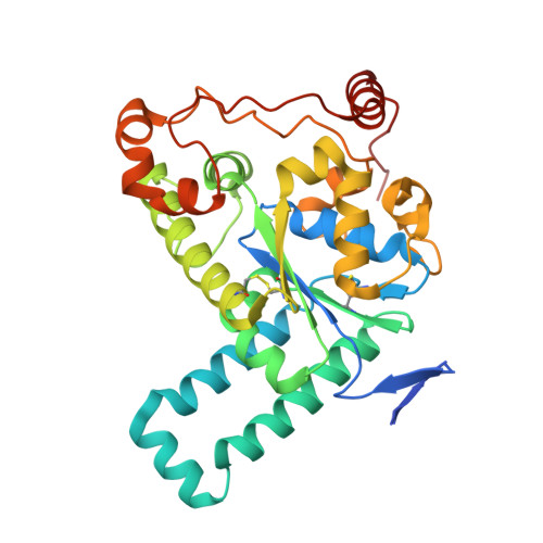
Crystal structure of human tyrosylprotein sulfotransferase 2 (TPST2) in cation-bound state


Experimental Data Snapshot

  • Method: X-RAY DIFFRACTION
  • Resolution: 1.75 Å
  • R-Value Free: 
    0.188 (Depositor), 0.185 (DCC) 
  • R-Value Work: 
    0.171 (Depositor), 0.170 (DCC) 
  • R-Value Observed: 
    0.172 (Depositor) 

Starting Model: experimental
View more details

wwPDB Validation 3D Report Full Report

Validation slider image for 9WWE

Ligand Structure Quality Assessment 


This is version 1.1 of the entry. See complete history

Literature

Structural characterization of metal binding in human tyrosylprotein sulfotransferase 2, TPST2.

Jin, M.Noh, C.Yang, J.Kim, H.Park, S.B.Kim, Y.C.Eom, S.H.

(2026) Sci Rep 16: 6066-6066

  • DOI: https://doi.org/10.1038/s41598-026-37189-4
  • Primary Citation Related Structures: 
    9WWE, 9WWF

  • PubMed Abstract: 

    Tyrosylprotein sulfotransferases (TPSTs) catalyze O-sulfation of tyrosine residues on secreted and membrane proteins, but the molecular basis for their stimulation by metal ions remains unclear. We determined the structures of the catalytic domain of human TPST2 with PAP and Na + (1.75 Å) or Mn 2+ (2.00 Å) bound and identified two conserved octahedral metal-binding sites. Anomalous diffraction at metal absorption edges confirmed the identity of the bound metals and demonstrated specific Mn 2+ binding. The Na + - and Mn 2+ -bound structures closely superimposed, suggesting activation without large conformational changes. Structural comparison with the apo structure and ensemble refinement revealed differences in local dynamics around the metal binding sites. The flexible α3-helix and α12-α13 loop in the apo structure were stabilized by Na + binding and further rigidified by Mn 2+ binding. These findings support an activation-by-ordering mechanism in which Na + binding generates a pre-activated state, with Mn 2+ subsequently establishing a catalytically competent ordering that lowers the entropic barrier at the active-site entrance. This framework reconciles longstanding biochemical observations and suggests that Mn 2+ availability within the Golgi can tune TPST2-dependent signaling.


  • Organizational Affiliation
    • Department of Life Sciences, Gwangju Institute of Science and Technology (GIST), Gwangju, Republic of Korea.

Macromolecule Content 

  • Total Structure Weight: 36.8 kDa 
  • Atom Count: 2,682 
  • Modeled Residue Count: 303 
  • Deposited Residue Count: 322 
  • Unique protein chains: 1

Macromolecules

Find similar proteins by:|  3D Structure
Entity ID: 1
MoleculeChains  Sequence LengthOrganismDetailsImage
Protein-tyrosine sulfotransferase 2322Homo sapiensMutation(s): 0 
Gene Names: TPST2
EC: 2.8.2.20
UniProt & NIH Common Fund Data Resources
Find proteins for O60704 (Homo sapiens)
Explore O60704 
Go to UniProtKB:  O60704
GTEx:  ENSG00000128294 
Entity Groups
Sequence Clusters30% Identity50% Identity70% Identity90% Identity95% Identity100% Identity
UniProt GroupO60704
Sequence Annotations
Expand
Reference Sequence

Small Molecules

Ligands 5 Unique
IDChains Name / Formula / InChI Key2D Diagram3D Interactions
A3P

Query on A3P


Download:Ideal Coordinates CCD File
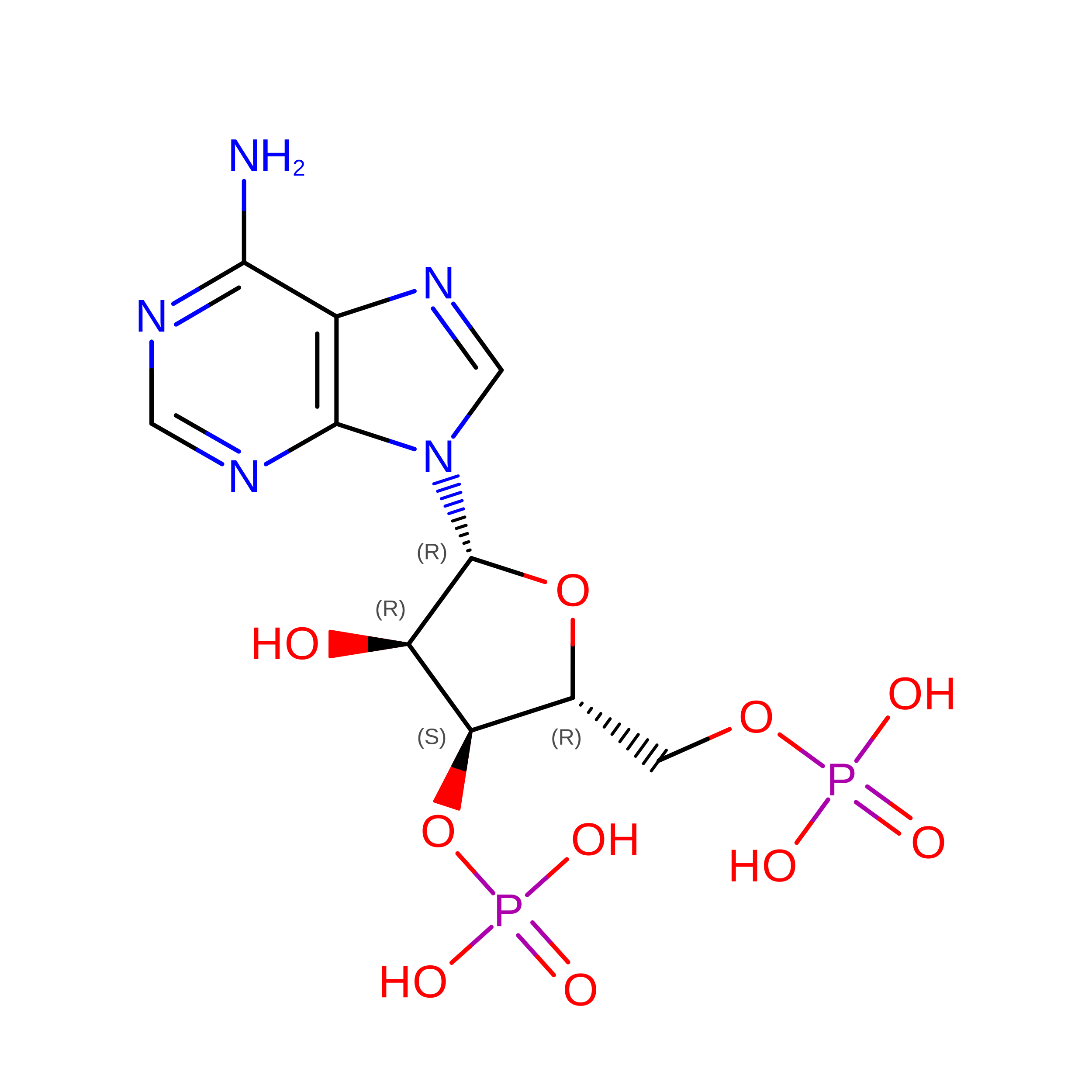
F [auth A]ADENOSINE-3'-5'-DIPHOSPHATE
C10 H15 N5 O10 P2
WHTCPDAXWFLDIH-KQYNXXCUSA-N
TLA
(Subject of Investigation/LOI)

Query on TLA


Download:Ideal Coordinates CCD File
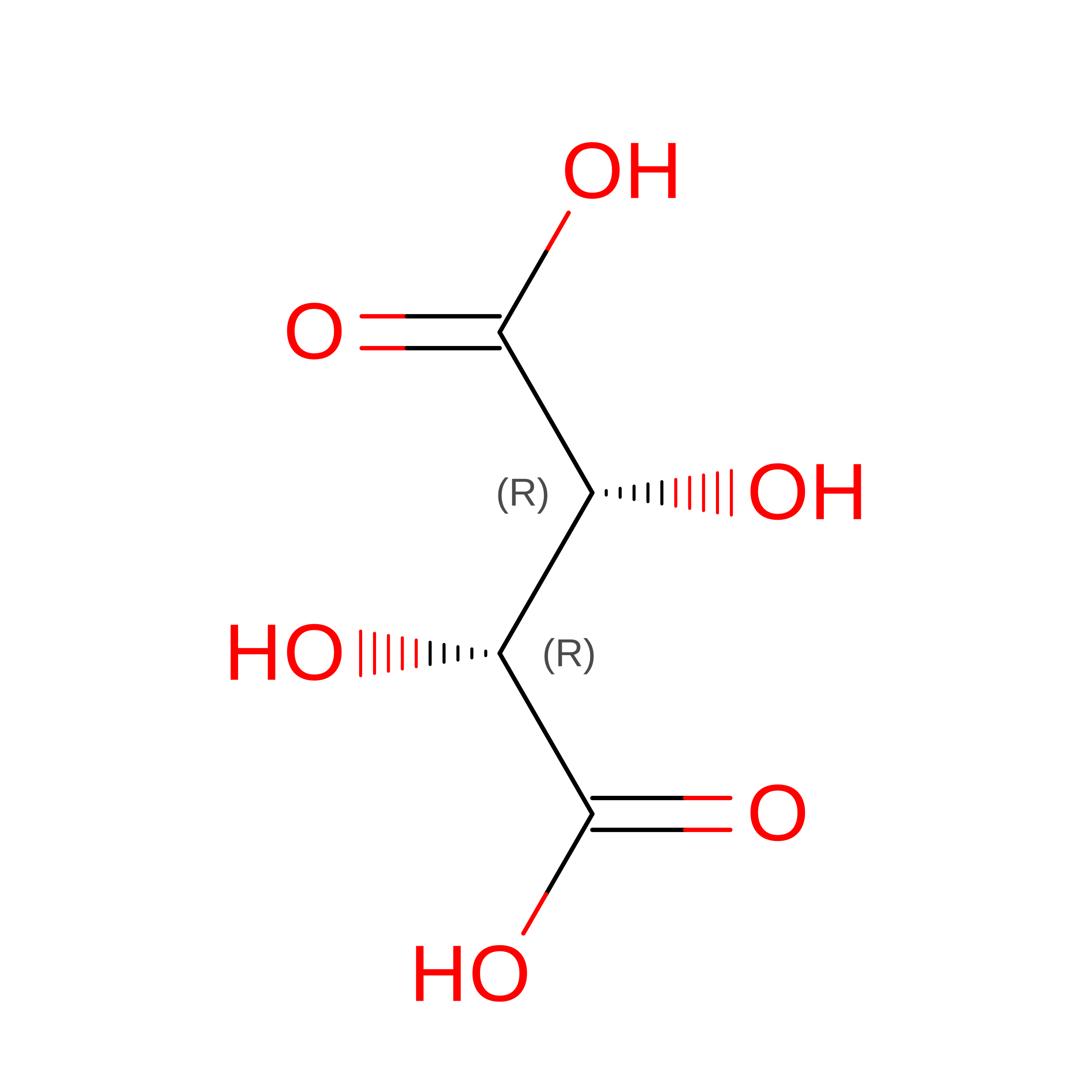
H [auth A]L(+)-TARTARIC ACID
C4 H6 O6
FEWJPZIEWOKRBE-JCYAYHJZSA-N
GOL

Query on GOL


Download:Ideal Coordinates CCD File
G [auth A]GLYCEROL
C3 H8 O3
PEDCQBHIVMGVHV-UHFFFAOYSA-N
ZN

Query on ZN


Download:Ideal Coordinates CCD File
D [auth A],
E [auth A]
ZINC ION
Zn
PTFCDOFLOPIGGS-UHFFFAOYSA-N
NA

Query on NA


Download:Ideal Coordinates CCD File
B [auth A],
C [auth A]
SODIUM ION
Na
FKNQFGJONOIPTF-UHFFFAOYSA-N

Experimental Data & Validation

Experimental Data

  • Method: X-RAY DIFFRACTION
  • Resolution: 1.75 Å
  • R-Value Free:  0.188 (Depositor), 0.185 (DCC) 
  • R-Value Work:  0.171 (Depositor), 0.170 (DCC) 
  • R-Value Observed: 0.172 (Depositor) 
Space Group: P 43 2 2
Unit Cell:
Length ( Å )Angle ( ˚ )
a = 102.254α = 90
b = 102.254β = 90
c = 103.6γ = 90
Software Package:
Software NamePurpose
XDSdata reduction
Aimlessdata scaling
PHENIXphasing
PHENIXrefinement
Cootmodel building

Structure Validation

View Full Validation Report


Ligand Structure Quality Assessment 


Entry History 

& Funding Information

Deposition Data


Funding OrganizationLocationGrant Number
National Research Foundation (NRF, Korea)Korea, Republic OfNRF-2021R1A2C1006267
National Research Foundation (NRF, Korea)Korea, Republic OfRS-2024-00344154
National Research Foundation (NRF, Korea)Korea, Republic OfRS-2024- 00440614

Revision History  (Full details and data files)

  • Version 1.0: 2025-12-31
    Type: Initial release
  • Version 1.1: 2026-07-15
    Changes: Database references