9VYG | pdb_00009vyg

Cyanopodophage Pan3 tail complex


Experimental Data Snapshot

  • Method: ELECTRON MICROSCOPY
  • Resolution: 3.50 Å
  • Aggregation State: PARTICLE 
  • Reconstruction Method: SINGLE PARTICLE 

wwPDB Validation   3D Report Full Report


This is version 1.1 of the entry. See complete history


Literature

Cryo-EM structure of cyanopodophage Pan3 reveals a modular tail architecture for host recognition.

Hou, P.Zhu, J.Yu, R.C.Yang, F.Du, K.Li, J.Kong, W.W.Wang, J.Chen, Y.Zhou, C.Z.Jiang, Y.L.

(2025) Structure 

  • DOI: https://doi.org/10.1016/j.str.2025.11.012
  • Primary Citation of Related Structures:  
    9VYE, 9VYF, 9VYG, 9VYH, 9VZI

  • PubMed Abstract: 

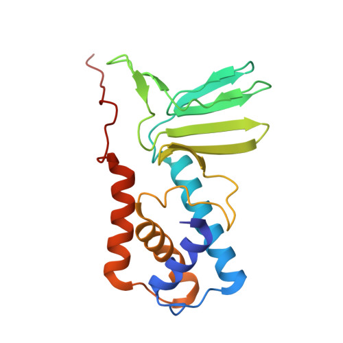
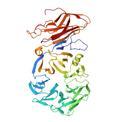
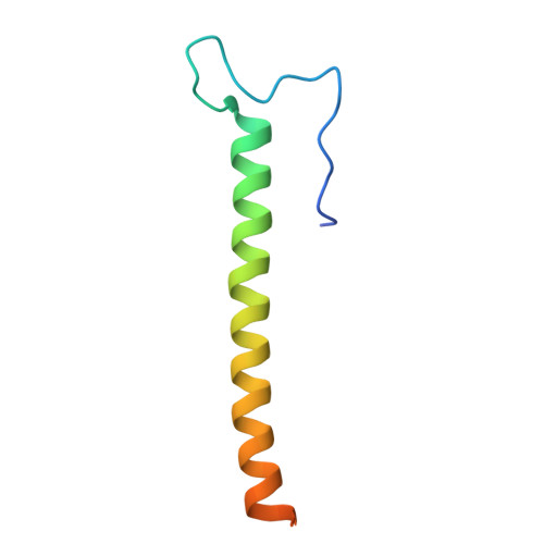
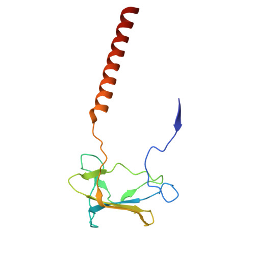
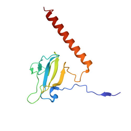
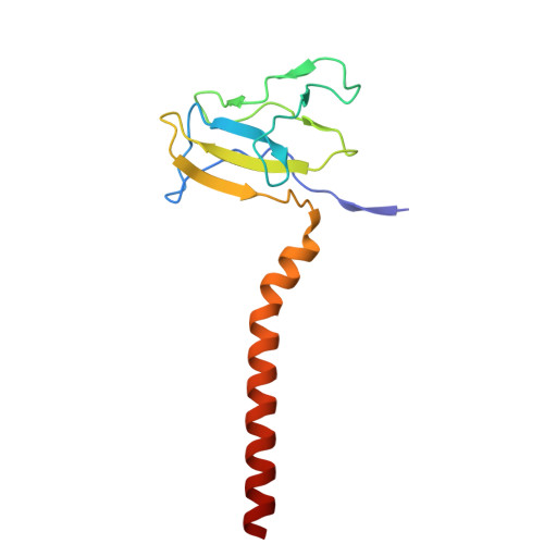
    Cyanophages, which are bacteriophages that specifically infect host cyanobacteria, also utilize the tail to initiate host recognition and adsorption. Owing to the limited structural information on cyanophages, our understanding of the mechanism by which cyanophages specifically recognize their hosts remains largely unknown. Here, we determined the intact cryoelectron microscopy structure of a freshwater cyanopodophage Pan3, which consists of an icosahedral shell and a short tail comprising four modular components: the dodecameric adaptor, hexameric nozzle, trimeric needle, and six heterohexameric tailspikes. Notably, each tailspike features an SGNH esterase domain fused to a lectin domain, forming a continuous groove complementary to the host lipopolysaccharide. These findings provide insights into the receptor engagement in Podoviridae, and establish a structural framework for cyanophage and host interactions that may guide future antibacterial interventions against harmful blooms.


  • Organizational Affiliation
    • Hefei National Research Center for Physical Sciences at the Microscale, Division of Life Sciences and Medicine, University of Science and Technology of China, Hefei, China.

Macromolecules
Find similar proteins by:  (by identity cutoff)  |  3D Structure
Entity ID: 1
MoleculeChains Sequence LengthOrganismDetailsImage
Adaptor protein
A, B, C, D, E
A, B, C, D, E, F, G, H, I, J, K, L
186Pseudanabaena phage Pan3Mutation(s): 0 
Entity Groups  
Sequence Clusters30% Identity50% Identity70% Identity90% Identity95% Identity100% Identity
Sequence Annotations
Expand
  • Reference Sequence
Find similar proteins by:  (by identity cutoff)  |  3D Structure
Entity ID: 2
MoleculeChains Sequence LengthOrganismDetailsImage
Stabilization protein
M, N, O, P, Q
M, N, O, P, Q, R
446Pseudanabaena phage Pan3Mutation(s): 0 
Entity Groups  
Sequence Clusters30% Identity50% Identity70% Identity90% Identity95% Identity100% Identity
Sequence Annotations
Expand
  • Reference Sequence
Find similar proteins by:  (by identity cutoff)  |  3D Structure
Entity ID: 3
MoleculeChains Sequence LengthOrganismDetailsImage
Needle protein
S, T, U
69Pseudanabaena phage Pan3Mutation(s): 0 
Entity Groups  
Sequence Clusters30% Identity50% Identity70% Identity90% Identity95% Identity100% Identity
Sequence Annotations
Expand
  • Reference Sequence
Find similar proteins by:  (by identity cutoff)  |  3D Structure
Entity ID: 4
MoleculeChains Sequence LengthOrganismDetailsImage
Tail spike proteinV [auth m]128Pseudanabaena phage Pan3Mutation(s): 0 
Entity Groups  
Sequence Clusters30% Identity50% Identity70% Identity90% Identity95% Identity100% Identity
Sequence Annotations
Expand
  • Reference Sequence
Find similar proteins by:  (by identity cutoff)  |  3D Structure
Entity ID: 5
MoleculeChains Sequence LengthOrganismDetailsImage
Tail spike proteinW [auth n]129Pseudanabaena phage Pan3Mutation(s): 0 
Entity Groups  
Sequence Clusters30% Identity50% Identity70% Identity90% Identity95% Identity100% Identity
Sequence Annotations
Expand
  • Reference Sequence
Find similar proteins by:  (by identity cutoff)  |  3D Structure
Entity ID: 6
MoleculeChains Sequence LengthOrganismDetailsImage
Tail spike proteinX [auth o]126Pseudanabaena phage Pan3Mutation(s): 0 
Entity Groups  
Sequence Clusters30% Identity50% Identity70% Identity90% Identity95% Identity100% Identity
Sequence Annotations
Expand
  • Reference Sequence
Experimental Data & Validation

Experimental Data

  • Method: ELECTRON MICROSCOPY
  • Resolution: 3.50 Å
  • Aggregation State: PARTICLE 
  • Reconstruction Method: SINGLE PARTICLE 
EM Software:
TaskSoftware PackageVersion
RECONSTRUCTIONRELION

Structure Validation

View Full Validation Report


Entry History & Funding Information

Deposition Data


Funding OrganizationLocationGrant Number
National Natural Science Foundation of China (NSFC)China32430001
National Natural Science Foundation of China (NSFC)China92451302

Revision History  (Full details and data files)

  • Version 1.0: 2025-11-26
    Type: Initial release
  • Version 1.1: 2026-01-28
    Changes: Data collection, Database references