9VDN | pdb_00009vdn

Cryo-EM structure of human ATP9A (AlF) E2P state open form


Experimental Data Snapshot

  • Method: ELECTRON MICROSCOPY
  • Resolution: 2.61 Å
  • Aggregation State: PARTICLE 
  • Reconstruction Method: SINGLE PARTICLE 

wwPDB Validation   3D Report Full Report


This is version 1.2 of the entry. See complete history


Literature

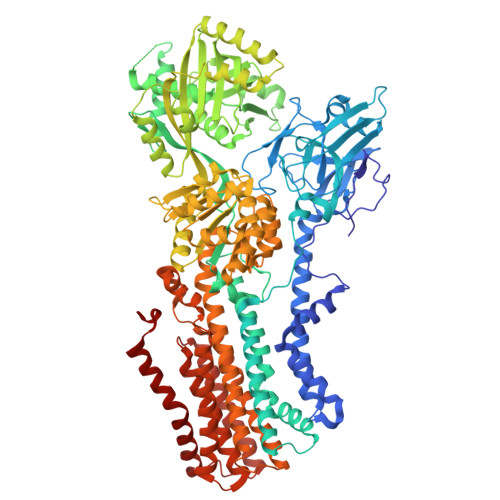
A unique gating mechanism revealed by the cryo-EM structure of monomeric ATP9A flippase.

Abe, K.Marimuthu, P.Qian, Y.Gopalasingam, C.C.Gerle, C.Shigematsu, H.Tanaka, K.Khandelia, H.

(2025) J Biological Chem 301: 110631-110631

  • DOI: https://doi.org/10.1016/j.jbc.2025.110631
  • Primary Citation of Related Structures:  
    9VDK, 9VDL, 9VDM, 9VDN

  • PubMed Abstract: 

    Among mammalian P4-ATPase flippases, only ATP9A and ATP9B do not require the auxiliary subunit CDC50 protein. Whilst its yeast homologue, Neo1, is essential for cell survival, little is known about mammalian ATP9A. We present cryo-EM structures of human monomeric ATP9A at a resolution reaching to 2.2Å, in the outward-facing E2P state. Two distinguishable conformations were obtained from a single sample, one with its outward gate open, and the other in its closed form. Unlike canonical gating observed for most P-type ATPases, which is driven by the movement of transmembrane (TM) helices 1 and 2 linked to the A domain, outward gating in ATP9A is achieved by the movement of TM6-10 helices, likely initiated by the unwinding of TM6. As a result, the volume of the phospholipid binding cavity in the open state surpasses that of other flippases, which could allow binding of phospholipids with larger hydrophilic head groups than that of phosphatidylserine. ATP9A shows an ATPase activity that is significantly increased by the addition of phospholipids that retain the overall negative charge, including phosphatidylserine, phosphatidylinositol and its phosphorylated species, compared to other electroneutral phospholipids. The observation of spontaneous binding of phosphorylated species of phosphatidylinositol in molecular simulation reinforces this fact. Our data provide mechanistic rationales for ATP9A gating, achieved by the rearrangement of the second half of the TM helices. Since TM4 - TM10 is anchored by the CDC50 protein subunit in other flippases, the here observed outward gating mechanism is unique to P4B-type flippases which function as a monomer.


  • Organizational Affiliation
    • Department of Chemistry, Faculty of Science, Hokkaido University, Japan. Electronic address: kabe@sci.hokudai.ac.jp.

Macromolecules
Find similar proteins by:  (by identity cutoff)  |  3D Structure
Entity ID: 1
MoleculeChains Sequence LengthOrganismDetailsImage
Probable phospholipid-transporting ATPase IIA1,015Homo sapiensMutation(s): 0 
Gene Names: ATP9AATPIIAKIAA0611
EC: 7.6.2.1
Membrane Entity: Yes 
UniProt & NIH Common Fund Data Resources
Find proteins for O75110 (Homo sapiens)
Explore O75110 
Go to UniProtKB:  O75110
PHAROS:  O75110
GTEx:  ENSG00000054793 
Entity Groups  
Sequence Clusters30% Identity50% Identity70% Identity90% Identity95% Identity100% Identity
UniProt GroupO75110
Sequence Annotations
Expand
  • Reference Sequence
Small Molecules
Ligands 5 Unique
IDChains Name / Formula / InChI Key2D Diagram3D Interactions
P5S (Subject of Investigation/LOI)
Query on P5S

Download Ideal Coordinates CCD File 
C [auth A]O-[(R)-{[(2R)-2,3-bis(octadecanoyloxy)propyl]oxy}(hydroxy)phosphoryl]-L-serine
C42 H82 N O10 P
TZCPCKNHXULUIY-RGULYWFUSA-N
PCW (Subject of Investigation/LOI)
Query on PCW

Download Ideal Coordinates CCD File 
D [auth A],
E [auth A],
G [auth A],
H [auth A]
1,2-DIOLEOYL-SN-GLYCERO-3-PHOSPHOCHOLINE
C44 H85 N O8 P
SNKAWJBJQDLSFF-NVKMUCNASA-O
CLR
Query on CLR

Download Ideal Coordinates CCD File 
F [auth A]CHOLESTEROL
C27 H46 O
HVYWMOMLDIMFJA-DPAQBDIFSA-N
ALF
Query on ALF

Download Ideal Coordinates CCD File 
I [auth A]TETRAFLUOROALUMINATE ION
Al F4
UYOMQIYKOOHAMK-UHFFFAOYSA-J
MG
Query on MG

Download Ideal Coordinates CCD File 
B [auth A]MAGNESIUM ION
Mg
JLVVSXFLKOJNIY-UHFFFAOYSA-N
Experimental Data & Validation

Experimental Data

  • Method: ELECTRON MICROSCOPY
  • Resolution: 2.61 Å
  • Aggregation State: PARTICLE 
  • Reconstruction Method: SINGLE PARTICLE 

Structure Validation

View Full Validation Report


Entry History & Funding Information

Deposition Data

  • Released Date: 2025-09-03 
  • Deposition Author(s): Abe, K.

Funding OrganizationLocationGrant Number
Japan Society for the Promotion of Science (JSPS)Japan24K01975

Revision History  (Full details and data files)

  • Version 1.0: 2025-09-03
    Type: Initial release
  • Version 1.1: 2025-09-10
    Changes: Data collection, Database references
  • Version 1.2: 2025-10-01
    Changes: Data collection, Database references