Design of Peptide Inhibitors Using Expression Tags: Structure of the Complex of Phosphopantetheine Adenylyltransferase with 17-Residue Expression-Tag Peptide and Citric Acid at 2.10 angstrom Resolution.
Ahmad, N., Kumar, V., Goel, V.K., Sharma, P., Sharma, S., Singh, T.P.(2025) Biochemistry 64: 4341-4353
- PubMed: 41069273 Search on PubMed
- DOI: https://doi.org/10.1021/acs.biochem.5c00465
- Primary Citation Related Structures:
9U54 - PubMed Abstract:
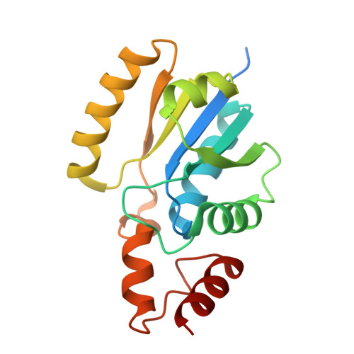
Phosphopantetheine adenylyltransferase (PPAT) catalyzes the transfer of an adenylyl group from adenosine triphosphate (ATP) to 4'-phosphopantetheine (PNS) to generate dephosphocoenzyme A (dPCoA) and pyrophosphate (PP i ). The dPCoA is required for the biosynthesis of coenzyme A (CoA), which is a vital cofactor in several essential biochemical reactions. PPAT enzyme from Enterobacter spp. ( Eb PPAT), cloned with a 30-residue-long N-terminal tag, was purified and crystallized. The structure determination of Eb PPAT revealed the presence of six protein molecules, A, B, C, D, E, and F, in the asymmetric unit, which formed three homodimers designated as A - B, C - D and E - F. At the N-termini of molecules B and F, 17 additional residues belonging to the expression tag were observed. These 17-residue segments of molecules B and F were located deep inside the PNS-binding sites of the adjacent molecules. In addition to this, six citric acid (CIT) molecules were observed in the ATP-binding sites of all six Eb PPAT molecules. Thus, the 17-mer peptide and CIT molecules filled the substrate-binding cleft of Eb PPAT completely. In order to estimate the binding affinity, the 17-mer tag peptide was synthesized. The K D value for the 17-mer peptide was found to be 1.7 × 10 -8 M. The K D value for the CIT molecule was 2.13 × 10 --5 M. These values indicated higher binding affinities of the 17-mer peptide and CIT molecule than those of the substrates, PNS and ATP, respectively. These results suggest that expression-tag fragments can be used to design the required peptide inhibitors of enzymes.
- Department of Biophysics, All India Institute of Medical Sciences, New Delhi 110029, India.
Organizational Affiliation: