9SRJ | pdb_00009srj

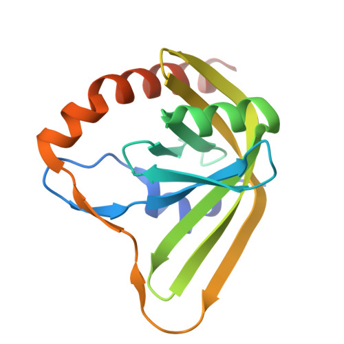
Prenylated FMN oxidative maturase PhdC, PEG-bound


Experimental Data Snapshot

  • Method: X-RAY DIFFRACTION
  • Resolution: 1.42 Å
  • R-Value Free: 
    0.204 (Depositor), 0.201 (DCC) 
  • R-Value Work: 
    0.181 (Depositor), 0.187 (DCC) 
  • R-Value Observed: 
    0.182 (Depositor) 

Starting Model: in silico
View more details

wwPDB Validation 3D Report Full Report

Validation slider image for 9SRJ

This is version 1.1 of the entry. See complete history

Literature

Structure and Mechanism of PhdC, a Prenylated-Flavin Maturase.

Whittall, D.R.Box, H.G.Payne, K.A.P.Marshall, S.A.Leys, D.

(2026) Proteins 94: 1019-1029

  • DOI: https://doi.org/10.1002/prot.70096
  • Primary Citation Related Structures: 
    9SRG, 9SRJ

  • PubMed Abstract: 

    Prenylated flavin mononucleotide (prFMN) is a modified flavin cofactor required by the UbiD family of (de)carboxylase enzymes. While the reduced prFMNH 2 form is produced by the flavin prenyltransferase UbiX, the corresponding two-electron oxidized prFMN iminium form is required to support UbiD catalysis. Thus, oxidative maturation of prFMNH 2 is required, which can be catalyzed by UbiD. However, heterologous (over)expression of UbiDs frequently leads to the accumulation of the stable but non-active one-electron oxidized purple prFMN radical species. A dedicated prFMN maturase enzyme (PhdC) from Mycolicibacterium fortuitum was recently identified as capable of catalyzing the oxidative maturation of prFMN radical to prFMN iminium , thereby enabling an effective supply of active cofactor to the associated phenazine-1-carboxylate (de)carboxylase PhdA. We report the crystal structure of PhdC in complex with flavin, revealing it is a distant member of the class I HpaC-like family of short-chain dimeric flavin reductases and demonstrate catalytic conversion of the prFMN radical species to prFMN iminium in the presence of oxygen or ferricyanide. Co-expression of PhdC or a distant homologue from Priestia megaterium (YclD) with the canonical UbiD from Escherichia coli leads to activation of the latter, similar in effect to co-expression with the prFMNH 2 -binding chaperone LpdD. Conserved Glu residues in the PhdC active site suggest catalysis occurs through C1' proton-abstraction coupled oxidation. This study thus provides both structural and mechanistic insight into the function of PhdC, adding to the expanding repertoire of prFMN-binding proteins associated with the widespread UbiDX system.


  • Organizational Affiliation
    • Manchester Institute of Biotechnology, University of Manchester, Manchester, UK.

Macromolecule Content 

  • Total Structure Weight: 37.04 kDa 
  • Atom Count: 2,582 
  • Modeled Residue Count: 302 
  • Deposited Residue Count: 326 
  • Unique protein chains: 1

Macromolecules

Find similar proteins by:|  3D Structure
Entity ID: 1
MoleculeChains  Sequence LengthOrganismDetailsImage
Pyridoxamine 5'-phosphate oxidase putative domain-containing protein
A, B
163Mycolicibacterium fortuitumMutation(s): 0 
Gene Names: XA26_16660
UniProt
Find proteins for A0A0N9XAG5 (Mycolicibacterium fortuitum)
Explore A0A0N9XAG5 
Go to UniProtKB:  A0A0N9XAG5
Entity Groups
Sequence Clusters30% Identity50% Identity70% Identity90% Identity95% Identity100% Identity
UniProt GroupA0A0N9XAG5
Sequence Annotations
Expand
Reference Sequence

Small Molecules

Ligands 3 Unique
IDChains Name / Formula / InChI Key2D Diagram3D Interactions
PGE

Query on PGE


Download:Ideal Coordinates CCD File
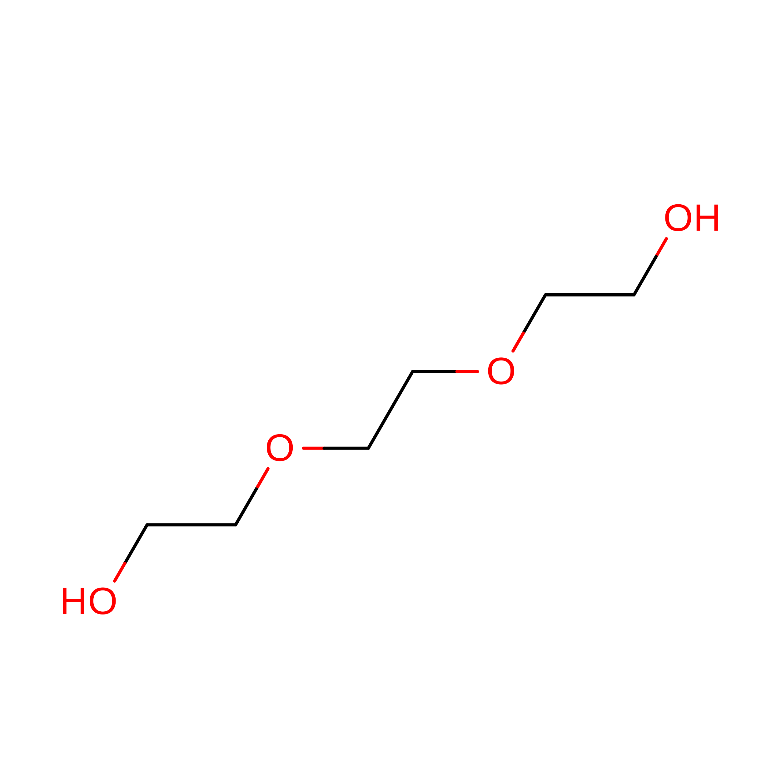
E [auth A]TRIETHYLENE GLYCOL
C6 H14 O4
ZIBGPFATKBEMQZ-UHFFFAOYSA-N
PEG

Query on PEG


Download:Ideal Coordinates CCD File
H [auth B],
I [auth B]
DI(HYDROXYETHYL)ETHER
C4 H10 O3
MTHSVFCYNBDYFN-UHFFFAOYSA-N
NA

Query on NA


Download:Ideal Coordinates CCD File
C [auth A],
D [auth A],
F [auth B],
G [auth B]
SODIUM ION
Na
FKNQFGJONOIPTF-UHFFFAOYSA-N

Experimental Data & Validation

Experimental Data

  • Method: X-RAY DIFFRACTION
  • Resolution: 1.42 Å
  • R-Value Free:  0.204 (Depositor), 0.201 (DCC) 
  • R-Value Work:  0.181 (Depositor), 0.187 (DCC) 
  • R-Value Observed: 0.182 (Depositor) 
Space Group: I 2 3
Unit Cell:
Length ( Å )Angle ( ˚ )
a = 121.253α = 90
b = 121.253β = 90
c = 121.253γ = 90
Software Package:
Software NamePurpose
PHENIXrefinement
PDB_EXTRACTdata extraction
Cootmodel building
AutoProcessdata scaling
PHENIXphasing

Structure Validation

View Full Validation Report


Entry History 

& Funding Information

Deposition Data


Funding OrganizationLocationGrant Number
Biotechnology and Biological Sciences Research Council (BBSRC)United Kingdom--

Revision History  (Full details and data files)

  • Version 1.0: 2025-12-17
    Type: Initial release
  • Version 1.1: 2026-04-15
    Changes: Database references