9SEB | pdb_00009seb

Crystal Structure of human exonuclease 1 (Exo1) with DNA and compound 20


Experimental Data Snapshot

  • Method: X-RAY DIFFRACTION
  • Resolution: 1.90 Å
  • R-Value Free: 
    0.275 (Depositor), 0.264 (DCC) 
  • R-Value Work: 
    0.236 (Depositor), 0.226 (DCC) 
  • R-Value Observed: 
    0.239 (Depositor) 

Starting Model: experimental
View more details

wwPDB Validation   3D Report Full Report


Ligand Structure Quality Assessment 


This is version 1.1 of the entry. See complete history


Literature

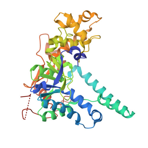
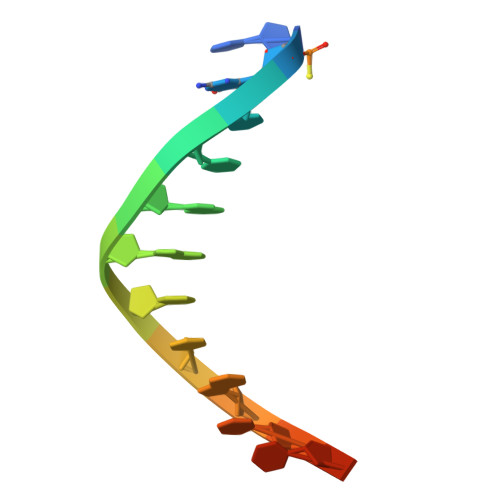
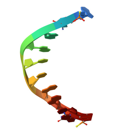
Discovery of ART5537: A Potent and Selective Small-Molecule Probe for EXO1.

Mann, S.E.Davis, O.A.Bomke, J.Cornaciu, I.Elinati, E.Follows, B.Galbiati, A.Geo, L.Grande, D.Jorand-Lebrun, C.Lademann, C.A.Lefranc, J.Leuthner, B.Mason, B.McWhirter, C.L.Musil, D.Pehl, U.Petersson, C.Pica, A.Pinto, M.F.Rajendra, E.Rakers, C.Rego, A.T.Robinson, H.M.R.Schwarz, D.Smith, G.C.M.Sorrell, F.J.Zenke, F.T.Heald, R.A.Burgdorf, L.T.

(2025) J Med Chem 68: 26432-26447

  • DOI: https://doi.org/10.1021/acs.jmedchem.5c02593
  • Primary Citation of Related Structures:  
    9SEB, 9SMO

  • PubMed Abstract: 

    Exonuclease 1 (EXO1) is emerging as a target of interest in oncology due to its involvement in multifaceted DNA metabolic processes, particularly in homologous recombination (HR). Evidence is building that BRCA1 -deficient cancers are sensitive to loss of EXO1, suggesting therapeutic potential for treating certain subsets of patients. However, EXO1 remains under-explored, with very few reported inhibitors, and there is a paucity of good quality, potent, and selective pharmacological tools to explore its biology. Here, we describe a metal-chelating fragment screen, which resulted in highly selective, submicromolar EXO1 hits. Our subsequent structure-based design and optimization led to the discovery of ART5537 , the first highly potent and selective EXO1 inhibitor. We demonstrate that inhibition of EXO1 leads to potent suppression of HR in cells and that the HR inhibition of ART5537 is driven exclusively by EXO1. Furthermore, we show that ART5537 sensitizes cancer cells to ionizing radiation (IR) and synergizes with PARP inhibitors (PARPi).


  • Organizational Affiliation
    • Artios Pharma Ltd., B940, Babraham Research Campus, Cambridge CB22 3FH, U.K.

Macromolecules

Find similar proteins by:  (by identity cutoff)  |  3D Structure
Entity ID: 3
MoleculeChains Sequence LengthOrganismDetailsImage
Exonuclease 1C [auth Z]358Homo sapiensMutation(s): 0 
Gene Names: EXO1EXOIHEX1
EC: 3.1
UniProt & NIH Common Fund Data Resources
Find proteins for Q9UQ84 (Homo sapiens)
Explore Q9UQ84 
Go to UniProtKB:  Q9UQ84
PHAROS:  Q9UQ84
GTEx:  ENSG00000174371 
Entity Groups  
Sequence Clusters30% Identity50% Identity70% Identity90% Identity95% Identity100% Identity
UniProt GroupQ9UQ84
Sequence Annotations
Expand
  • Reference Sequence

Find similar nucleic acids by:  Sequence   |   3D Structure  

Entity ID: 1
MoleculeChains LengthOrganismImage
DNA (5'-D(*CP*(GS)P*CP*TP*AP*GP*TP*CP*GP*TP*CP*AP*(PST))-3')13synthetic construct
Sequence Annotations
Expand
  • Reference Sequence

Find similar nucleic acids by:  Sequence   |   3D Structure  

Entity ID: 2
MoleculeChains LengthOrganismImage
DNA (5'-D(P*AP*(SC)P*GP*AP*CP*TP*AP*GP*CP*(GS))-3')10synthetic construct
Sequence Annotations
Expand
  • Reference Sequence
Small Molecules
Ligands 4 Unique
IDChains Name / Formula / InChI Key2D Diagram3D Interactions
A1JNC (Subject of Investigation/LOI)
Query on A1JNC

Download Ideal Coordinates CCD File 
G [auth Z]1-(5-chloranyl-3-methyl-pyridin-2-yl)-5-oxidanyl-3-[(1~{S})-1-phenylethyl]-2~{H}-pyrido[2,1-f][1,2,4]triazine-4,6-dione
C21 H19 Cl N4 O3
PABWYWJUARAIGO-AWEZNQCLSA-N
FMT
Query on FMT

Download Ideal Coordinates CCD File 
H [auth Z],
I [auth Z]
FORMIC ACID
C H2 O2
BDAGIHXWWSANSR-UHFFFAOYSA-N
MG
Query on MG

Download Ideal Coordinates CCD File 
D [auth Z],
E [auth Z]
MAGNESIUM ION
Mg
JLVVSXFLKOJNIY-UHFFFAOYSA-N
NA
Query on NA

Download Ideal Coordinates CCD File 
F [auth Z]SODIUM ION
Na
FKNQFGJONOIPTF-UHFFFAOYSA-N
Experimental Data & Validation

Experimental Data

  • Method: X-RAY DIFFRACTION
  • Resolution: 1.90 Å
  • R-Value Free:  0.275 (Depositor), 0.264 (DCC) 
  • R-Value Work:  0.236 (Depositor), 0.226 (DCC) 
  • R-Value Observed: 0.239 (Depositor) 
Space Group: P 43 21 2
Unit Cell:
Length ( Å )Angle ( ˚ )
a = 69.836α = 90
b = 69.836β = 90
c = 181.661γ = 90
Software Package:
Software NamePurpose
BUSTERrefinement
autoPROCdata reduction
autoPROCdata scaling
PHASERphasing

Structure Validation

View Full Validation Report


Ligand Structure Quality Assessment 


Entry History & Funding Information

Deposition Data


Funding OrganizationLocationGrant Number
Other privateUnited Kingdom--
Other privateGermany--

Revision History  (Full details and data files)

  • Version 1.0: 2025-12-17
    Type: Initial release
  • Version 1.1: 2026-01-07
    Changes: Database references