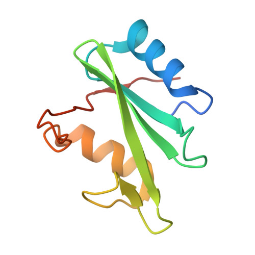
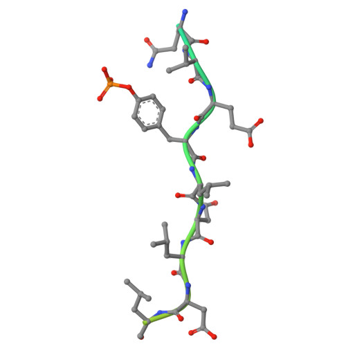
The non-receptor tyrosine phosphatase SHP2 (SH2 domain-containing protein tyrosine phosphatase 2) (PTPN11) is a regulator of diverse cellular functions including mitogenic activation and cell migration. It comprises two tandem Src-homology 2 (SH2) domains followed by the catalytic domain and is autoinhibited by the N-terminal SH2 domain blocking access to the active site. Mutations influencing auto-inhibition have been implicated in cancer and other diseases, and allosteric inhibitors have been developed that stabilize the inactive state. Here, we show that the intrinsically disordered bis-phosphorylated SHP2-activating peptide pY 627 pY 659 -Gab1 binds to both SH2 domains, undergoing partial ordering in the process. In addition to eliciting changes in SH2 domain dynamics, the peptide reorganizes their relative orientations to generate a new SH2-SH2 interface. Our data suggest an active conformation for SHP2 that is also applicable to the hematopoietic cell-specific SHP1 (PTPN6), shedding light on the activation mechanism of both enzymes and paving the way for the development of novel compounds to modulate SHP2 activity.
Organizational Affiliation:
Institut für Biochemie und Biotechnologie, Martin-Luther-Universität Halle-Wittenberg, 06120 Halle (Saale), Germany; Institut für Molekulare Medizin, Martin-Luther-Universität Halle-Wittenberg, 06120 Halle (Saale), Germany; Charles-Tanford-Proteinzentrum, Martin-Luther-Universität Halle-Wittenberg, 06120 Halle (Saale), Germany.
Institut für Biochemie und Biotechnologie, Martin-Luther-Universität Halle-Wittenberg, 06120 Halle (Saale), Germany; Charles-Tanford-Proteinzentrum, Martin-Luther-Universität Halle-Wittenberg, 06120 Halle (Saale), Germany.
Institut für Biochemie und Biotechnologie, Martin-Luther-Universität Halle-Wittenberg, 06120 Halle (Saale), Germany; ZIK HALOmem, Biozentrum, Martin-Luther-Universität Halle-Wittenberg, 06120 Halle (Saale), Germany; Institute of Chemical Biology, National Hellenic Research Foundation, 11635 Athens, Greece.
Institut für Physik, Martin-Luther-Universität Halle-Wittenberg, 06120 Halle (Saale), Germany; ZIK HALOmem, Biozentrum, Martin-Luther-Universität Halle-Wittenberg, 06120 Halle (Saale), Germany; Mitteldeutsches Zentrum für Struktur und Dynamik der Proteine, Martin-Luther-Universität Halle-Wittenberg, 06120 Halle (Saale), Germany.
Institut für Biochemie und Biotechnologie, Martin-Luther-Universität Halle-Wittenberg, 06120 Halle (Saale), Germany; ZIK HALOmem, Biozentrum, Martin-Luther-Universität Halle-Wittenberg, 06120 Halle (Saale), Germany; Institute of Chemical Biology, National Hellenic Research Foundation, 11635 Athens, Greece; Mitteldeutsches Zentrum für Struktur und Dynamik der Proteine, Martin-Luther-Universität Halle-Wittenberg, 06120 Halle (Saale), Germany.