Fluorinated HIV-1 protease inhibitors containing chiral hydroxyethylbenzene and indanol as P2' ligands with potent activity against drug-resistant variants.
Kaur, J., Spielvogel, E., Nageswara Rao, D., Rusere, L.N., Shaqra, A.M., Lockbaum, G.J., Maryam, A., Yilmaz, N.K., Swanstrom, R., Schiffer, C.A., Ali, A.(2025) Eur J Med Chem 304: 118510-118510
- PubMed: 41448052 Search on PubMed
- DOI: https://doi.org/10.1016/j.ejmech.2025.118510
- Primary Citation Related Structures:
9PYX, 9PZ1, 9Q0T, 9Q13, 9Q1C, 9Q1P, 9Q3P, 9Q3T, 9Q5D, 9YKP, 9YRA, 9YRR, 9YRY - PubMed Abstract:
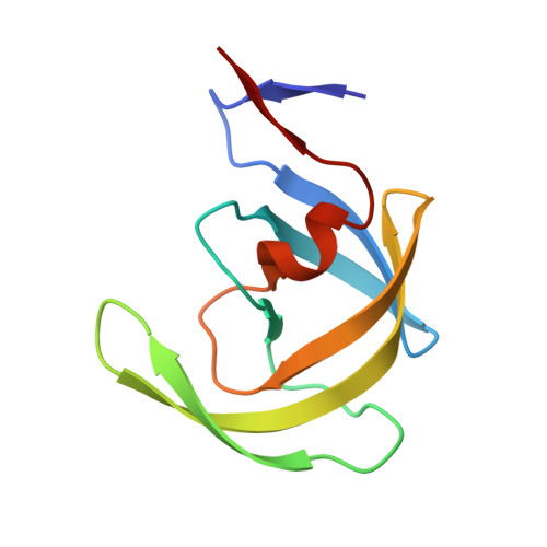
HIV-1 protease inhibitors are potent antiretroviral drugs, but their efficacy is often undermined by poor pharmacokinetics and drug resistance. Here, we employed a structure-guided design strategy to improve the potency and resistance profile of HIV-1 protease inhibitors by optimizing hydrogen bonding and van der Waals interactions within the protease substrate envelope. A series of darunavir analogs were designed by incorporating chiral 4-(1-hydroxyethyl)benzene and 1-indanol moieties as P2' ligands, in combination with P1 fluorination. The resulting compounds showed distinct potency profiles depending on the conformational flexibility of the P2' hydroxyl group. Notably, the P1 fluorinated compounds exhibited excellent antiviral potency against highly drug-resistant HIV-1 variants. Analysis of the protease-inhibitor cocrystal structures revealed that, similar to the 4-(1-hydroxyethyl)benzene moiety, both stereoisomers of the 1-indanol moiety make direct hydrogen bonding interactions with the backbone NH of Asp30'. To maintain polar interactions in the S2' subsite of HIV-1 protease, the orientation of the (R)-indanol moiety was flipped relative to the (S)-1-indanol moiety. The SAR data and structural analysis offer insights for further optimization to improve potency against drug-resistant HIV-1 variants.
- Department of Biochemistry and Molecular Biotechnology, University of Massachusetts Chan Medical School, Worcester, MA, 01605, United States.
Organizational Affiliation: