9P1T | pdb_00009p1t

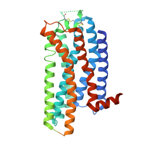
A2AR-BRIL in complex with ZM241385


Experimental Data Snapshot

  • Method: ELECTRON MICROSCOPY
  • Resolution: 3.15 Å
  • Aggregation State: PARTICLE 
  • Reconstruction Method: SINGLE PARTICLE 

wwPDB Validation 3D Report Full Report

Validation slider image for 9P1T

This is version 1.1 of the entry. See complete history

Literature

Systematic metabolite screening identifies functional regulators of the adenosine A2A receptor.

Rao, P.Rathinaswamy, M.Chan, M.Paredes, A.G.Patel, C.Wranik, B.J.Powell, J.Eaton, D.Rutter, J.Hicks, K.G.Mafi, A.Hao, Q.

(2025) Commun Chem 9: 19-19

  • DOI: https://doi.org/10.1038/s42004-025-01825-3
  • Primary Citation Related Structures: 
    9P1S, 9P1T

  • PubMed Abstract: 

    The adenosine A2A receptor (A2AR) is a Class A G protein-coupled receptor (GPCR) that regulates inflammation, glucose metabolism, and energy homeostasis in metabolically active tissues. While the effects of small-molecule ligands and protein interactions with A2AR have been extensively studied, the regulatory influence of endogenous metabolites remains unexplored. To address this gap, we employed the Mass spectrometry Integrated with equilibrium Dialysis for the discovery of Allostery Systematically (MIDAS) platform to screen a library of human metabolites for interactions with A2AR. This approach identified 180 metabolites that interact with A2AR, including allosteric and orthosteric modulators. We characterized the mechanisms of three metabolites previously unreported to interact with A2AR: prostaglandin D2, an allosteric antagonist that fully inhibits receptor signaling, and two orthosteric agonists, S-adenosyl-L-homocysteine and 2'-deoxyadenosine, that fully activate A2AR. Overall, these findings highlight the potential of the MIDAS platform to uncover previously unrecognized metabolite-GPCR interactions for research and therapeutic applications.


  • Organizational Affiliation
    • Calico Life Sciences LLC, 1170 Veterans Blvd, South San Francisco, CA, USA.

Macromolecule Content 

  • Total Structure Weight: 49.15 kDa 
  • Atom Count: 2,179 
  • Modeled Residue Count: 271 
  • Deposited Residue Count: 430 
  • Unique protein chains: 1

Macromolecules

Find similar proteins by:|  3D Structure
Entity ID: 1
MoleculeChains  Sequence LengthOrganismDetailsImage
Adenosine receptor A2a,Soluble cytochrome b562430Homo sapiensMutation(s): 12 
Gene Names: ADORA2AADORA2cybC
UniProt & NIH Common Fund Data Resources
Find proteins for P29274 (Homo sapiens)
Explore P29274 
Go to UniProtKB:  P29274
PHAROS:  P29274
GTEx:  ENSG00000128271 
Entity Groups
Sequence Clusters30% Identity50% Identity70% Identity90% Identity95% Identity100% Identity
UniProt GroupP29274
Sequence Annotations
Expand
Reference Sequence

Small Molecules

Ligands 2 Unique
IDChains Name / Formula / InChI Key2D Diagram3D Interactions
LMN

Query on LMN


Download:Ideal Coordinates CCD File
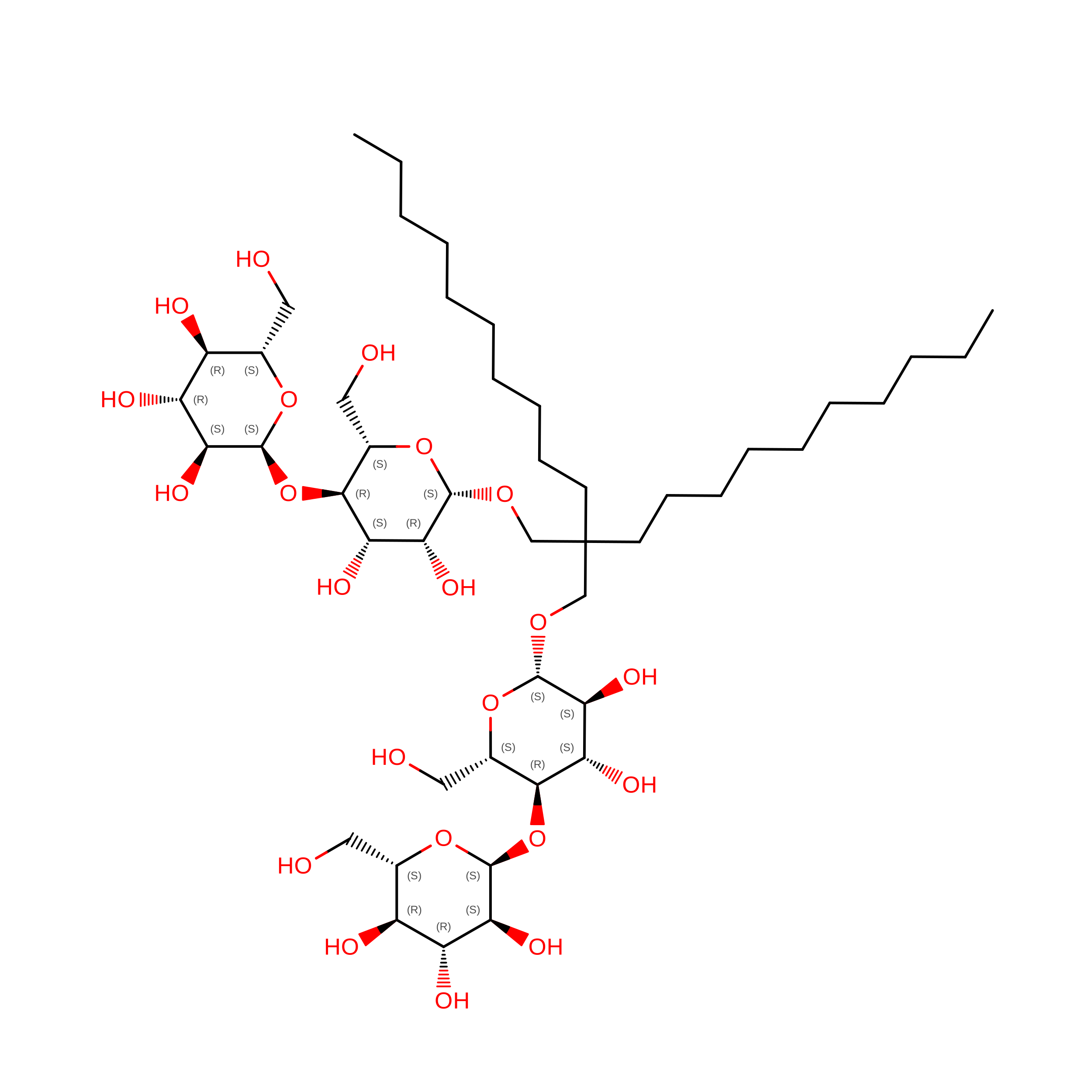
B [auth A]Lauryl Maltose Neopentyl Glycol
C47 H88 O22
MADJBYLAYPCCOO-XYPZXBMFSA-N
ZMA

Query on ZMA


Download:Ideal Coordinates CCD File
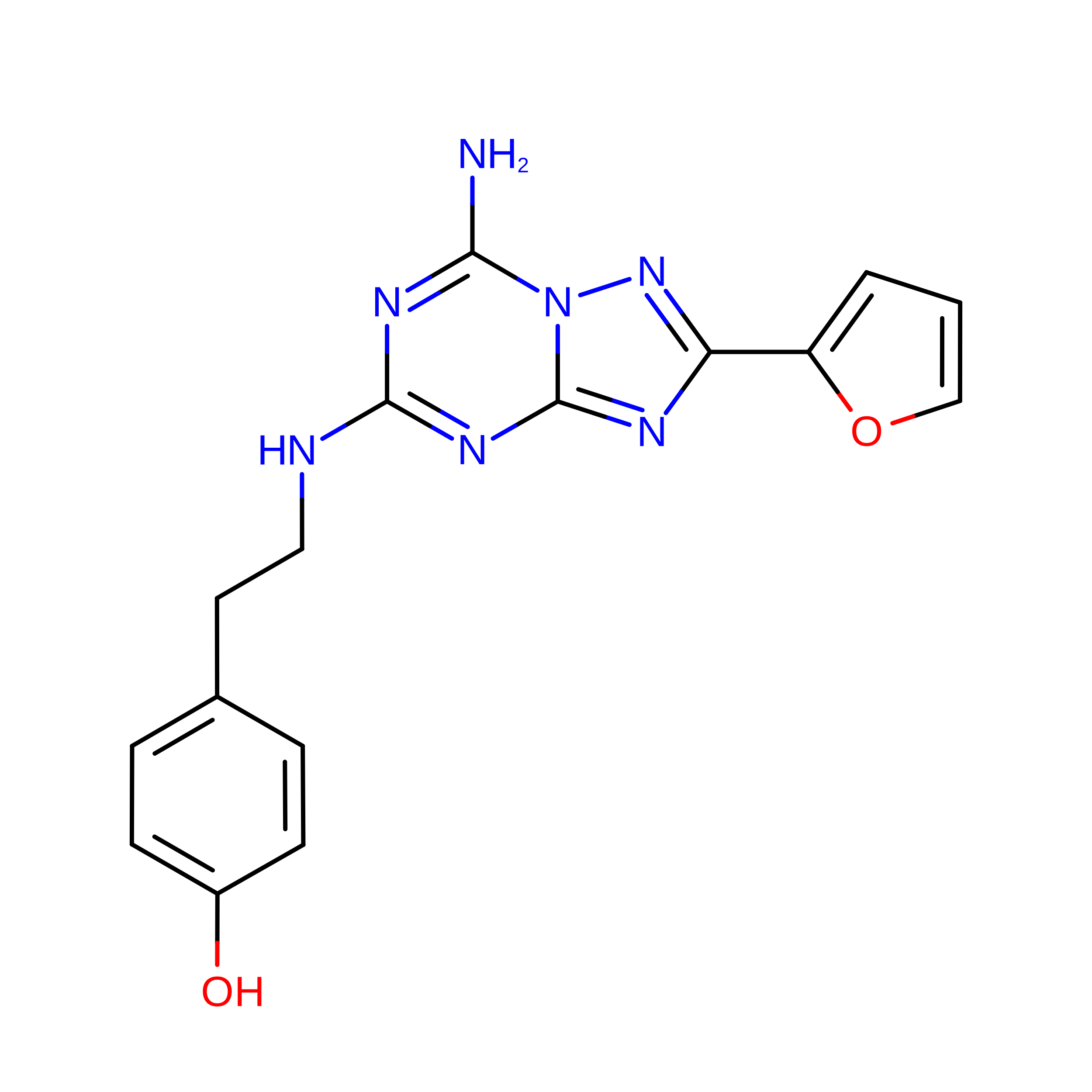
C [auth A]4-{2-[(7-amino-2-furan-2-yl[1,2,4]triazolo[1,5-a][1,3,5]triazin-5-yl)amino]ethyl}phenol
C16 H15 N7 O2
PWTBZOIUWZOPFT-UHFFFAOYSA-N

Experimental Data & Validation

Experimental Data

  • Method: ELECTRON MICROSCOPY
  • Resolution: 3.15 Å
  • Aggregation State: PARTICLE 
  • Reconstruction Method: SINGLE PARTICLE 
EM Software:
TaskSoftware PackageVersion
MODEL REFINEMENTPHENIX1.19.2_4158
RECONSTRUCTIONRELION

Structure Validation

View Full Validation Report


Entry History 

& Funding Information

Deposition Data


Funding OrganizationLocationGrant Number
Not fundedUnited States--

Revision History  (Full details and data files)

  • Version 1.0: 2025-12-24
    Type: Initial release
  • Version 1.1: 2026-01-28
    Changes: Data collection, Database references