9OXQ | pdb_00009oxq

X-ray crystal structure of Beta-Barrel forming peptide macrocycle

  • Classification: DE NOVO PROTEIN
  • Organism(s): synthetic construct
  • Mutation(s): No 

  • Deposited: 2025-06-04 Released: 2025-06-18 
  • Deposition Author(s): Dang, V.T., Nguyen, A.
  • Funding Organization(s): National Institutes of Health/National Institute of General Medical Sciences (NIH/NIGMS)

Experimental Data Snapshot

  • Method: X-RAY DIFFRACTION
  • Resolution: 1.55 Å
  • R-Value Free: 
    0.191 (Depositor), 0.191 (DCC) 
  • R-Value Work: 
    0.193 (Depositor), 0.194 (DCC) 
  • R-Value Observed: 
    0.193 (Depositor) 

Starting Model: experimental
View more details

wwPDB Validation   3D Report Full Report


This is version 1.2 of the entry. See complete history


Literature

beta-barrels from short macrocyclic peptides.

Dang, V.T.Martynowycz, M.W.McElheny, D.Nguyen, A.I.

(2026) Chem Commun (Camb) 

  • DOI: https://doi.org/10.1039/d5cc06640a
  • Primary Citation of Related Structures:  
    9OXP, 9OXQ, 9Q63, 9Q6E

  • PubMed Abstract: 

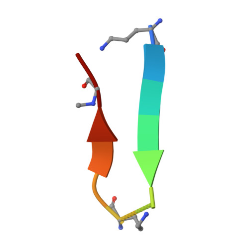
    β-Barrels are ubiquitous motifs in protein structures, but the fundamental rules underlying their formation are unclear, and their de novo design remains highly challenging. Small peptides that form barrels are especially scarce. Here, we report barrels with the shortest staves (6 residues, ∼60% of previous record) and smallest shear number ( S = 4) so far, formed from 12-residue macrocyclic peptides. The miniature barrel has anomalous structural features, demonstrated by solution phase and crystallographic characterization; there is a pronounced and essential backbone kink imparted by an achiral residue, N -methylglycine, as well as four structural water molecules stitching the seams of the barrel. These results provide insights into how extremely short sequences could form barrel assemblies.


  • Organizational Affiliation
    • Department of Chemistry, University of Illinois Chicago, Chicago, Illinois 60607, USA. andyn@uic.edu.

Macromolecules

Find similar proteins by:  Sequence   |   3D Structure  

Entity ID: 1
MoleculeChains Sequence LengthOrganismDetailsImage
MC4H (ORN)WFHSH(ORN)FIH(SAR)H peptide macrocycle
A, C, E, G
12synthetic constructMutation(s): 0 
Entity Groups  
Sequence Clusters30% Identity50% Identity70% Identity90% Identity95% Identity100% Identity
Sequence Annotations
Expand
  • Reference Sequence

Find similar proteins by:  Sequence   |   3D Structure  

Entity ID: 2
MoleculeChains Sequence LengthOrganismDetailsImage
MC4H (ORN)FIH(SAR)H(ORN)WFHSH peptide macrocycle
B, D, F, H
12synthetic constructMutation(s): 0 
Entity Groups  
Sequence Clusters30% Identity50% Identity70% Identity90% Identity95% Identity100% Identity
Sequence Annotations
Expand
  • Reference Sequence
Experimental Data & Validation

Experimental Data

  • Method: X-RAY DIFFRACTION
  • Resolution: 1.55 Å
  • R-Value Free:  0.191 (Depositor), 0.191 (DCC) 
  • R-Value Work:  0.193 (Depositor), 0.194 (DCC) 
  • R-Value Observed: 0.193 (Depositor) 
Space Group: P 1 21 1
Unit Cell:
Length ( Å )Angle ( ˚ )
a = 25.012α = 90
b = 40.715β = 103.41
c = 46.939γ = 90
Software Package:
Software NamePurpose
PHENIXrefinement
XDSdata reduction
XDSdata scaling
PHENIXphasing

Structure Validation

View Full Validation Report


Entry History & Funding Information

Deposition Data


Funding OrganizationLocationGrant Number
National Institutes of Health/National Institute of General Medical Sciences (NIH/NIGMS)United States1R35GM154793-01

Revision History  (Full details and data files)

  • Version 1.0: 2025-06-18
    Type: Initial release
  • Version 1.1: 2025-07-09
    Changes: Structure summary
  • Version 1.2: 2026-01-14
    Changes: Database references