9OO1 | pdb_00009oo1

Influenza A Virus Group 2 Hemagglutinin (H7, Strain SH13) in Complex with a Potent Small-Molecule Entry Inhibitor ING-16-36


Experimental Data Snapshot

  • Method: ELECTRON MICROSCOPY
  • Resolution: 2.76 Å
  • Aggregation State: PARTICLE 
  • Reconstruction Method: SINGLE PARTICLE 

Starting Model: experimental
View more details

wwPDB Validation 3D Report Full Report

Validation slider image for 9OO1

This is version 1.1 of the entry. See complete history

Literature

Mechanistic insights into the small-molecule inhibition of influenza A virus entry.

Xu, Y.Anirudhan, V.Gaisina, I.N.Du, H.Alqarni, S.Moore, T.W.Caffrey, M.Manicassamy, B.Zhou, T.Rong, L.Xu, K.

(2025) Proc Natl Acad Sci U S A 122: e2503899122-e2503899122

  • DOI: https://doi.org/10.1073/pnas.2503899122
  • Primary Citation Related Structures: 
    9ONZ, 9OO1

  • PubMed Abstract: 

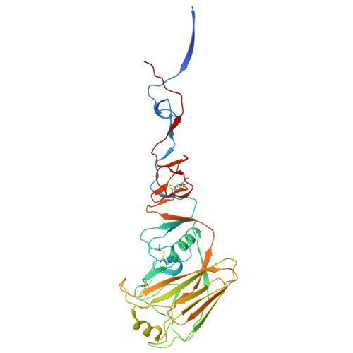
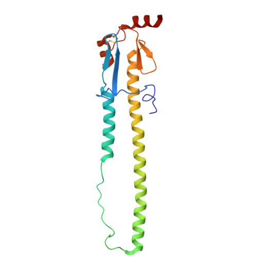
    Influenza A virus (IAV) is a zoonotic pathogen responsible for seasonal and pandemic flu. The extensive genetic and antigenic diversity within and between IAV phylogenetic groups presents major challenges for developing universal vaccines and broad-spectrum antiviral therapies. Current interventions provide limited protection due to the virus's high mutation rate and capacity for immune evasion. Recent advancements in viral hemagglutinin (HA)-targeting small-molecule entry inhibitors offer a promising avenue to overcome these limitations. Here, we present structural and functional analyses of two group 2 HA-specific small-molecule inhibitors recently identified by our team. Cryogenic electron microscopy (cryo-EM) structures revealed that these inhibitors bind a conserved pocket within the HA stalk, likely interfering with the conformational rearrangements necessary for membrane fusion and viral entry. Structure-guided mutagenesis confirmed the critical roles of key interacting residues and uncovered distinct resistance profiles between the two compounds, as well as in comparison to Arbidol, a previously reported HA inhibitor. Notably, our structural analysis highlights intrinsic barriers to achieving cross-group inhibition with current small-molecule designs. To address this, we propose an alternative strategy for broadening antiviral coverage. Together, these findings provide mechanistic insights into IAV entry inhibition and a foundation for the rational design of next-generation anti-influenza therapeutics.


  • Organizational Affiliation
    • Department of Veterinary Biosciences, The Ohio State University, Columbus, OH 43210.

Macromolecule Content 

  • Total Structure Weight: 174.8 kDa 
  • Atom Count: 11,775 
  • Modeled Residue Count: 1,470 
  • Deposited Residue Count: 1,530 
  • Unique protein chains: 2

Macromolecules

Find similar proteins by:|  3D Structure
Entity ID: 1
MoleculeChains  Sequence LengthOrganismDetailsImage
Hemagglutinin HA1
A, B, C
338Influenza A virusMutation(s): 0 
Gene Names: HA
UniProt
Find proteins for A0A8E4VQE2 (Influenza A virus)
Explore A0A8E4VQE2 
Go to UniProtKB:  A0A8E4VQE2
Entity Groups
Sequence Clusters30% Identity50% Identity70% Identity90% Identity95% Identity100% Identity
UniProt GroupA0A8E4VQE2
Glycosylation
Glycosylation Sites: 1
Sequence Annotations
Expand
Reference Sequence
Find similar proteins by:|  3D Structure
Entity ID: 2
MoleculeChains  Sequence LengthOrganismDetailsImage
Hemagglutinin HA2
D, E, F
172Influenza A virusMutation(s): 0 
Gene Names: HA
UniProt
Find proteins for A0A8E4QKM0 (Influenza A virus)
Explore A0A8E4QKM0 
Go to UniProtKB:  A0A8E4QKM0
Entity Groups
Sequence Clusters30% Identity50% Identity70% Identity90% Identity95% Identity100% Identity
UniProt GroupA0A8E4QKM0
Glycosylation
Glycosylation Sites: 2
Sequence Annotations
Expand
Reference Sequence

Oligosaccharides

Help  
Entity ID: 3
MoleculeChains Length2D Diagram GlycosylationD Interactions
beta-D-mannopyranose-(1-4)-2-acetamido-2-deoxy-beta-D-glucopyranose-(1-4)-2-acetamido-2-deoxy-beta-D-glucopyranose
G, H, I
3N-Glycosylation
Glycosylation Resources
GlyTouCan: G15407YE
GlyCosmos: G15407YE
GlyGen: G15407YE

Small Molecules

Ligands 2 Unique
IDChains Name / Formula / InChI Key2D Diagram3D Interactions
A1CC3
(Subject of Investigation/LOI)

Query on A1CC3


Download:Ideal Coordinates CCD File
N [auth B],
Q [auth C],
S [auth D]
(2M,4S)-2-(2-chloropyridin-4-yl)-N-cyclohexyl-5,7-dimethylimidazo[1,2-a]pyrimidin-3-amine
C19 H22 Cl N5
HERNWKUOCFEFTO-UHFFFAOYSA-N
NAG

Query on NAG


Download:Ideal Coordinates CCD File
J [auth A]
K [auth A]
L [auth B]
M [auth B]
O [auth C]
J [auth A],
K [auth A],
L [auth B],
M [auth B],
O [auth C],
P [auth C],
R [auth D],
T [auth E],
U [auth F]
2-acetamido-2-deoxy-beta-D-glucopyranose
C8 H15 N O6
OVRNDRQMDRJTHS-FMDGEEDCSA-N

Experimental Data & Validation

Experimental Data

  • Method: ELECTRON MICROSCOPY
  • Resolution: 2.76 Å
  • Aggregation State: PARTICLE 
  • Reconstruction Method: SINGLE PARTICLE 
EM Software:
TaskSoftware PackageVersion
MODEL REFINEMENTPHENIX
RECONSTRUCTIONcryoSPARC

Structure Validation

View Full Validation Report


Entry History 

& Funding Information

Deposition Data

  • Released Date: 2025-07-16 
  • Deposition Author(s): Xu, Y., Xu, K.

Funding OrganizationLocationGrant Number
Not fundedUnited States--

Revision History  (Full details and data files)

  • Version 1.0: 2025-07-16
    Type: Initial release
  • Version 1.1: 2025-08-27
    Changes: Data collection, Database references