Building molecular model series from heterogeneous CryoEM structures using Gaussian mixture models and deep neural networks.
Chen, M.(2025) Commun Biol 8: 798-798
- PubMed: 40415012 Search on PubMedSearch on PubMed Central
- DOI: https://doi.org/10.1038/s42003-025-08202-9
- Primary Citation Related Structures:
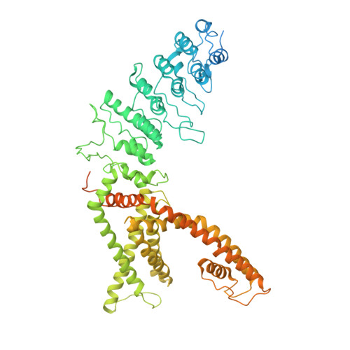
9OGK - PubMed Abstract:
Cryogenic electron microscopy (CryoEM) produces structures of macromolecules at near-atomic resolution. However, building molecular models with good stereochemical geometry from those structures can be challenging and time-consuming, especially when many structures are obtained from datasets with conformational heterogeneity. Here we present a model refinement protocol that automatically generates series of molecular models from CryoEM datasets, which describe the dynamics of the macromolecular system and have near-perfect geometry scores. This method makes it easier to interpret the movement of the protein complex from heterogeneity analysis and to compare the structural dynamics observed from CryoEM data with results from other experimental and simulation techniques.
- Division of CryoEM and Bioimaging, SSRL, SLAC National Accelerator Laboratory, Stanford University, Menlo Park, CA, USA. muyuanc@stanford.edu.
Organizational Affiliation: