5IRX

Structure of TRPV1 in complex with DkTx and RTX, determined in lipid nanodisc


Experimental Data Snapshot

  • Method: ELECTRON MICROSCOPY
  • Resolution: 2.95 Å
  • Aggregation State: PARTICLE 
  • Reconstruction Method: SINGLE PARTICLE 

wwPDB Validation   3D Report Full Report


This is version 1.4 of the entry. See complete history


Literature

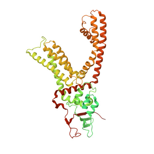
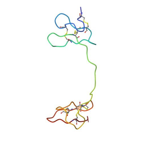
TRPV1 structures in nanodiscs reveal mechanisms of ligand and lipid action.

Gao, Y.Cao, E.Julius, D.Cheng, Y.

(2016) Nature 534: 347-351

  • DOI: https://doi.org/10.1038/nature17964
  • Primary Citation of Related Structures:  
    5IRX, 5IRZ, 5IS0

  • PubMed Abstract: 

    When integral membrane proteins are visualized in detergents or other artificial systems, an important layer of information is lost regarding lipid interactions and their effects on protein structure. This is especially relevant to proteins for which lipids have both structural and regulatory roles. Here we demonstrate the power of combining electron cryo-microscopy with lipid nanodisc technology to ascertain the structure of the rat TRPV1 ion channel in a native bilayer environment. Using this approach, we determined the locations of annular and regulatory lipids and showed that specific phospholipid interactions enhance binding of a spider toxin to TRPV1 through formation of a tripartite complex. Furthermore, phosphatidylinositol lipids occupy the binding site for capsaicin and other vanilloid ligands, suggesting a mechanism whereby chemical or thermal stimuli elicit channel activation by promoting the release of bioactive lipids from a critical allosteric regulatory site.


  • Organizational Affiliation

    Department of Physiology, University of California, San Francisco, California 94143, USA.


Macromolecules
Find similar proteins by:  (by identity cutoff)  |  3D Structure
Entity ID: 1
MoleculeChains Sequence LengthOrganismDetailsImage
Transient receptor potential cation channel subfamily V member 1
A, B, C, D
636Rattus norvegicusMutation(s): 0 
Gene Names: Trpv1Vr1Vr1l
Membrane Entity: Yes 
UniProt
Find proteins for O35433 (Rattus norvegicus)
Explore O35433 
Go to UniProtKB:  O35433
Entity Groups  
Sequence Clusters30% Identity50% Identity70% Identity90% Identity95% Identity100% Identity
UniProt GroupO35433
Sequence Annotations
Expand
  • Reference Sequence
Find similar proteins by:  (by identity cutoff)  |  3D Structure
Entity ID: 2
MoleculeChains Sequence LengthOrganismDetailsImage
Tau-theraphotoxin-Hs1a
E, F
75Cyriopagopus schmidtiMutation(s): 0 
UniProt
Find proteins for P0CH43 (Cyriopagopus schmidti)
Explore P0CH43 
Go to UniProtKB:  P0CH43
Entity Groups  
Sequence Clusters30% Identity50% Identity70% Identity90% Identity95% Identity100% Identity
UniProt GroupP0CH43
Sequence Annotations
Expand
  • Reference Sequence
Small Molecules
Ligands 4 Unique
IDChains Name / Formula / InChI Key2D Diagram3D Interactions
6EU
Query on 6EU

Download Ideal Coordinates CCD File 
J [auth A],
M [auth B],
P [auth C],
U [auth D]
resiniferatoxin
C37 H40 O9
DSDNAKHZNJAGHN-MXTYGGKSSA-N
6O8
Query on 6O8

Download Ideal Coordinates CCD File 
G [auth A],
K [auth B],
N [auth C],
Q [auth D]
(4R,7S)-4-hydroxy-N,N,N-trimethyl-4,9-dioxo-7-[(pentanoyloxy)methyl]-3,5,8-trioxa-4lambda~5~-phosphatetradecan-1-aminium
C19 H39 N O8 P
IZTOQGOABQJQQN-KRWDZBQOSA-O
6OE
Query on 6OE

Download Ideal Coordinates CCD File 
H [auth A]
I [auth A]
L [auth B]
O [auth C]
S [auth D]
H [auth A],
I [auth A],
L [auth B],
O [auth C],
S [auth D],
T [auth D],
W [auth E],
Z [auth F]
(2S)-3-{[(S)-(2-aminoethoxy)(hydroxy)phosphoryl]oxy}-2-(hexanoyloxy)propyl hexanoate
C17 H34 N O8 P
PELYUHWUVHDSSU-HNNXBMFYSA-N
6O9
Query on 6O9

Download Ideal Coordinates CCD File 
R [auth D],
V [auth E],
X [auth F],
Y [auth F]
(2S)-2-(acetyloxy)-3-{[(R)-(2-aminoethoxy)(hydroxy)phosphoryl]oxy}propyl pentanoate
C12 H24 N O8 P
PLBNSCVUDWCBFD-NSHDSACASA-N
Binding Affinity Annotations 
IDSourceBinding Affinity
6EU BindingDB:  5IRX Ki: min: 0.13, max: 23 (nM) from 3 assay(s)
IC50: 180 (nM) from 1 assay(s)
Experimental Data & Validation

Experimental Data

  • Method: ELECTRON MICROSCOPY
  • Resolution: 2.95 Å
  • Aggregation State: PARTICLE 
  • Reconstruction Method: SINGLE PARTICLE 
EM Software:
TaskSoftware PackageVersion
RECONSTRUCTIONRELION1.3
MODEL REFINEMENTCoot
MODEL REFINEMENTPHENIX

Structure Validation

View Full Validation Report


Entry History & Funding Information

Deposition Data


Funding OrganizationLocationGrant Number
National Institutes of Health/National Institute of Neurological Disorders and Stroke (NIH/NINDS)United StatesR01NS047723
National Institutes of Health/National Institute of Neurological Disorders and Stroke (NIH/NINDS)United StatesR37NS065071
National Institutes of Health/Office of the DirectorUnited StatesS10OD020054
National Institutes of Health/National Institute of General Medical Sciences (NIH/NIGMS)United StatesR01GM098672

Revision History  (Full details and data files)

  • Version 1.0: 2016-05-25
    Type: Initial release
  • Version 1.1: 2016-06-22
    Changes: Database references
  • Version 1.2: 2016-11-30
    Changes: Source and taxonomy
  • Version 1.3: 2017-09-13
    Changes: Author supporting evidence, Data collection
  • Version 1.4: 2019-12-18
    Changes: Author supporting evidence