9NUE | pdb_00009nue

Y20S after Mg2+ (Sec18) - Class 3


Experimental Data Snapshot

  • Method: ELECTRON MICROSCOPY
  • Resolution: 3.36 Å
  • Aggregation State: PARTICLE 
  • Reconstruction Method: SINGLE PARTICLE 

wwPDB Validation   3D Report Full Report


This is version 1.0 of the entry. See complete history


Literature

SNARE disassembly requires Sec18/NSF side loading.

Khan, Y.A.White, K.I.Pfuetzner, R.A.Singal, B.Esquivies, L.Mckenzie, G.Liu, F.DeLong, K.Choi, U.B.Montabana, E.Mclaughlin, T.Wickner, W.T.Brunger, A.T.

(2025) Nat Struct Mol Biol 32: 1708-1720

  • DOI: https://doi.org/10.1038/s41594-025-01590-w
  • Primary Citation of Related Structures:  
    9CRU, 9CRX, 9N22, 9NG2, 9NLU, 9NLW, 9NLY, 9NLZ, 9NM1, 9NUD, 9NUE, 9NUZ, 9NV0, 9NV1, 9NV9, 9NVD

  • PubMed Abstract: 

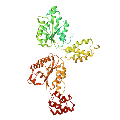
    SNARE (soluble N-ethylmaleimide-sensitive factor (NSF) attachment protein receptor) proteins drive membrane fusion at different cell compartments as their core domains zipper into a parallel four-helix bundle. After fusion, these bundles are disassembled by the AAA+ (ATPase associated with diverse cellular activities) protein Sec18/NSF and its adaptor Sec17/α-SNAP to make them available for subsequent rounds of membrane fusion. SNARE domains are often flanked by C-terminal transmembrane or N-terminal domains. Previous structures of the NSF-α-SNAP-SNARE complex revealed binding to the D1 ATPase pore, posing a topological constraint as SNARE transmembrane domains would prevent complete substrate threading as suggested for other AAA+ systems. Using mass spectrometry in yeast cells, we show N-terminal SNARE domain interactions with Sec18, exacerbating this topological issue. We present cryo-electron microscopy (cryo-EM) structures of a yeast SNARE complex, Sec18 and Sec17 in a nonhydrolyzing condition, which show SNARE Sso1 threaded through the D1 and D2 ATPase rings of Sec18, with its folded, N-terminal Habc domain interacting with the D2 ring. This domain does not unfold during Sec18/NSF activity. Cryo-EM structures under hydrolyzing conditions revealed substrate-released and substrate-free states of Sec18 with a coordinated opening in the side of the ATPase rings. Thus, Sec18/NSF operates by substrate side loading and unloading topologically constrained SNARE substrates.


  • Organizational Affiliation
    • Department of Molecular and Cellular Physiology, Stanford University, Stanford, CA, USA. yousuf@stanford.edu.

Macromolecules
Find similar proteins by:  (by identity cutoff)  |  3D Structure
Entity ID: 1
MoleculeChains Sequence LengthOrganismDetailsImage
Vesicular-fusion protein SEC18
A, B, C, D, E
A, B, C, D, E, F, G
761Saccharomyces cerevisiaeMutation(s): 0 
Gene Names: SEC18YBR080CYBR0736
UniProt
Find proteins for P18759 (Saccharomyces cerevisiae (strain ATCC 204508 / S288c))
Explore P18759 
Go to UniProtKB:  P18759
Entity Groups  
Sequence Clusters30% Identity50% Identity70% Identity90% Identity95% Identity100% Identity
UniProt GroupP18759
Sequence Annotations
Expand
  • Reference Sequence
Small Molecules
Ligands 3 Unique
IDChains Name / Formula / InChI Key2D Diagram3D Interactions
ATP (Subject of Investigation/LOI)
Query on ATP

Download Ideal Coordinates CCD File 
AA [auth G]
J [auth A]
M [auth B]
P [auth C]
S [auth D]
AA [auth G],
J [auth A],
M [auth B],
P [auth C],
S [auth D],
V [auth E],
Y [auth F]
ADENOSINE-5'-TRIPHOSPHATE
C10 H16 N5 O13 P3
ZKHQWZAMYRWXGA-KQYNXXCUSA-N
ADP (Subject of Investigation/LOI)
Query on ADP

Download Ideal Coordinates CCD File 
I [auth A]
L [auth B]
O [auth C]
R [auth D]
U [auth E]
I [auth A],
L [auth B],
O [auth C],
R [auth D],
U [auth E],
X [auth F]
ADENOSINE-5'-DIPHOSPHATE
C10 H15 N5 O10 P2
XTWYTFMLZFPYCI-KQYNXXCUSA-N
MG (Subject of Investigation/LOI)
Query on MG

Download Ideal Coordinates CCD File 
H [auth A]
K [auth B]
N [auth C]
Q [auth D]
T [auth E]
H [auth A],
K [auth B],
N [auth C],
Q [auth D],
T [auth E],
W [auth F],
Z [auth G]
MAGNESIUM ION
Mg
JLVVSXFLKOJNIY-UHFFFAOYSA-N
Experimental Data & Validation

Experimental Data

  • Method: ELECTRON MICROSCOPY
  • Resolution: 3.36 Å
  • Aggregation State: PARTICLE 
  • Reconstruction Method: SINGLE PARTICLE 

Structure Validation

View Full Validation Report


Entry History & Funding Information

Deposition Data


Funding OrganizationLocationGrant Number
National Institutes of Health/National Institute of Mental Health (NIH/NIMH)United States5R37MH063105
National Institutes of Health/National Institute of Mental Health (NIH/NIMH)United States1F31MH134477

Revision History  (Full details and data files)

  • Version 1.0: 2025-10-01
    Type: Initial release