9NCS | pdb_00009ncs

RNase A in complex with Uridine Vanadate and decavanadates


Experimental Data Snapshot

  • Method: X-RAY DIFFRACTION
  • Resolution: 1.83 Å
  • R-Value Free: 
    0.209 (Depositor), 0.216 (DCC) 
  • R-Value Work: 
    0.177 (Depositor), 0.187 (DCC) 
  • R-Value Observed: 
    0.181 (Depositor) 

Starting Model: experimental
View more details

wwPDB Validation 3D Report Full Report

Validation slider image for 9NCS

Ligand Structure Quality Assessment 


This is version 1.1 of the entry. See complete history

Literature

Pseudouridine residues as substrates for serum ribonucleases.

Gutierrez, C.S.Silkenath, B.Kojasoy, V.Pich, J.A.Lim, D.C.Raines, R.T.

(2025) RNA 31: 1542-1556

  • DOI: https://doi.org/10.1261/rna.080404.125
  • Primary Citation Related Structures: 
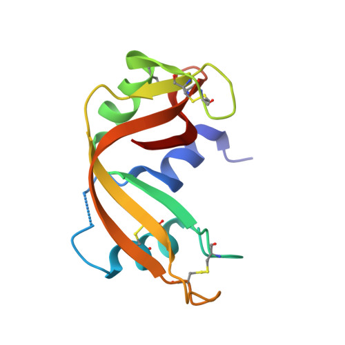
    9NCS, 9O4V, 9O5B

  • PubMed Abstract: 

    In clinical uses, RNA must maintain its integrity in serum that contains ribonucleases (RNases), especially RNase 1, which is a human homolog of RNase A. These omnipresent enzymes catalyze the cleavage of the P-O 5'' bond on the 3' side of pyrimidine residues. Pseudouridine (Ψ) is the most abundant modified nucleoside in natural RNA. The substitution of uridine (U) with Ψ or N 1 ‑methylpseudouridine (m 1 Ψ) reduces the immunogenicity of mRNA and increases ribosomal translation, and these modified nucleosides are key components of RNA-based vaccines. Here, we assessed the ability of RNase A and RNase 1 to catalyze the cleavage of the P-O 5'' bond on the 3' side of Ψ and m 1 Ψ. We find that these enzymes catalyze the cleavage of UpA up to 10‑fold more efficiently than the cleavage of ΨpA or m 1 ΨpA. X-ray crystallography of enzyme-bound nucleoside 2',3'‑cyclic vanadate complexes and molecular dynamics simulations of enzyme·dinucleotide complexes show that U, Ψ, and m 1 Ψ bind to RNase A and RNase 1 in a similar manner. Quantum chemistry calculations suggested that the higher reactivity of UpA is intrinsic, arising from an inductive effect that decreases the p K a of the 2'‑hydroxy group of U and enhances its nucleophilicity toward the P-O 5'' bond. Experimentally, we found that UpA does indeed undergo spontaneous hydrolysis faster than does m 1 ΨpA. Our findings inform the continuing development of RNA-based vaccines and therapeutic agents.


  • Organizational Affiliation
    • Massachusetts Institute of Technology.

Macromolecule Content 

  • Total Structure Weight: 31.56 kDa 
  • Atom Count: 2,323 
  • Modeled Residue Count: 243 
  • Deposited Residue Count: 248 
  • Unique protein chains: 1

Macromolecules

Find similar proteins by:|  3D Structure
Entity ID: 1
MoleculeChains  Sequence LengthOrganismDetailsImage
Ribonuclease pancreatic
A, B
124Bos taurusMutation(s): 0 
EC: 4.6.1.18
UniProt
Find proteins for P61823 (Bos taurus)
Explore P61823 
Go to UniProtKB:  P61823
Entity Groups
Sequence Clusters30% Identity50% Identity70% Identity90% Identity95% Identity100% Identity
UniProt GroupP61823
Sequence Annotations
Expand
Reference Sequence

Experimental Data & Validation

Experimental Data

  • Method: X-RAY DIFFRACTION
  • Resolution: 1.83 Å
  • R-Value Free:  0.209 (Depositor), 0.216 (DCC) 
  • R-Value Work:  0.177 (Depositor), 0.187 (DCC) 
  • R-Value Observed: 0.181 (Depositor) 
Space Group: C 1 2 1
Unit Cell:
Length ( Å )Angle ( ˚ )
a = 100.053α = 90
b = 32.677β = 90.36
c = 72.327γ = 90
Software Package:
Software NamePurpose
PHENIXrefinement
XDSdata scaling
XDSdata reduction
PHASERphasing

Structure Validation

View Full Validation Report


Ligand Structure Quality Assessment 


Entry History 

& Funding Information

Deposition Data


Funding OrganizationLocationGrant Number
National Institutes of Health/National Cancer Institute (NIH/NCI)United StatesR01 CA073808

Revision History  (Full details and data files)

  • Version 1.0: 2025-09-24
    Type: Initial release
  • Version 1.1: 2025-10-29
    Changes: Database references