9N4L | pdb_00009n4l

Composite map for GluK2 in the apo state with asymmetric ligand binding domain


Experimental Data Snapshot

  • Method: ELECTRON MICROSCOPY
  • Resolution: 4.24 Å
  • Aggregation State: PARTICLE 
  • Reconstruction Method: SINGLE PARTICLE 

wwPDB Validation 3D Report Full Report

Validation slider image for 9N4L

This is version 1.1 of the entry. See complete history

Literature

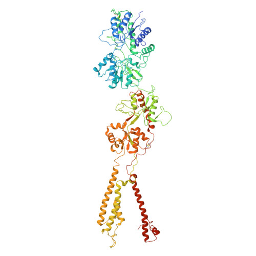
Activation of kainate receptor GluK2-Neto2 complex.

Gangwar, S.P.Yelshanskaya, M.V.Yen, L.Y.Newton, T.P.Sobolevsky, A.I.

(2025) Nat Struct Mol Biol 32: 2176-2184

  • DOI: https://doi.org/10.1038/s41594-025-01656-9
  • Primary Citation Related Structures: 
    9N4L, 9N4M, 9N4N, 9N4O, 9N4P, 9N4Q, 9N4R, 9N4S, 9N4T

  • PubMed Abstract: 

    Kainate receptors (KARs) are tetrameric, ligand-gated ion channels of the ionotropic glutamate receptor family that mediate excitatory neurotransmission and modulate neuronal circuits and synaptic plasticity during development of the central nervous system. KARs are implicated in psychiatric and neurological diseases and represent a target of therapeutic intervention. Native KARs form complexes with neuropilin and tolloid-like auxiliary subunits (Neto1 and Neto2), which modulate their function, trafficking and synaptic localization. Here we present structures of rat GluK2 KAR in the apo closed state and in the open states activated by agonist kainate and positive allosteric modulator BPAM344, solved in the presence and absence of Neto2 using time-resolved cryo-electron microscopy. While the binding of Neto2 does not change the behavior of individual or dimeric ligand-binding domains (LBDs) or the ion channel, it prevents tightening of the interface between two LBD dimers during activation and slows the kinetics of deactivation. Our structures illuminate the mechanism of KAR activation and its modulation by Neto2.


  • Organizational Affiliation
    • Department of Biochemistry and Molecular Biophysics, Columbia University, New York, NY, USA.

Macromolecule Content 

  • Total Structure Weight: 418.86 kDa 
  • Atom Count: 27,183 
  • Modeled Residue Count: 3,354 
  • Deposited Residue Count: 3,632 
  • Unique protein chains: 1

Macromolecules

Find similar proteins by:|  3D Structure
Entity ID: 1
MoleculeChains  Sequence LengthOrganismDetailsImage
Glutamate receptor ionotropic, kainate 2
A, B, C, D
908Rattus norvegicusMutation(s): 0 
Gene Names: Grik2Glur6
Membrane Entity: Yes 
UniProt
Find proteins for P42260 (Rattus norvegicus)
Explore P42260 
Go to UniProtKB:  P42260
Entity Groups
Sequence Clusters30% Identity50% Identity70% Identity90% Identity95% Identity100% Identity
UniProt GroupP42260
Glycosylation
Glycosylation Sites: 6Go to GlyGen: P42260-1
Sequence Annotations
Expand
Reference Sequence

Oligosaccharides

Help  
Entity ID: 2
MoleculeChains Length2D Diagram GlycosylationD Interactions
2-acetamido-2-deoxy-beta-D-glucopyranose-(1-4)-2-acetamido-2-deoxy-beta-D-glucopyranose
E, G, I, J, L
E, G, I, J, L, M
2N-Glycosylation
Glycosylation Resources
GlyTouCan: G42666HT
GlyCosmos: G42666HT
GlyGen: G42666HT
Entity ID: 3
MoleculeChains Length2D Diagram GlycosylationD Interactions
alpha-D-mannopyranose-(1-3)-[alpha-D-mannopyranose-(1-6)]beta-D-mannopyranose-(1-3)-2-acetamido-2-deoxy-beta-D-glucopyranose-(1-4)-2-acetamido-2-deoxy-beta-D-glucopyranose
F
5N-Glycosylation
Entity ID: 4
MoleculeChains Length2D Diagram GlycosylationD Interactions
alpha-D-mannopyranose-(1-3)-[alpha-D-mannopyranose-(1-6)]beta-D-mannopyranose-(1-4)-2-acetamido-2-deoxy-beta-D-glucopyranose-(1-4)-2-acetamido-2-deoxy-beta-D-glucopyranose
H, K
5N-Glycosylation
Glycosylation Resources
GlyTouCan: G22768VO
GlyCosmos: G22768VO
GlyGen: G22768VO

Small Molecules

Ligands 1 Unique
IDChains Name / Formula / InChI Key2D Diagram3D Interactions
NAG

Query on NAG


Download:Ideal Coordinates CCD File
AA [auth D]
BA [auth D]
CA [auth D]
N [auth A]
O [auth A]
AA [auth D],
BA [auth D],
CA [auth D],
N [auth A],
O [auth A],
P [auth A],
Q [auth A],
R [auth A],
S [auth B],
T [auth B],
U [auth B],
V [auth C],
W [auth C],
X [auth C],
Y [auth D],
Z [auth D]
2-acetamido-2-deoxy-beta-D-glucopyranose
C8 H15 N O6
OVRNDRQMDRJTHS-FMDGEEDCSA-N

Experimental Data & Validation

Experimental Data

  • Method: ELECTRON MICROSCOPY
  • Resolution: 4.24 Å
  • Aggregation State: PARTICLE 
  • Reconstruction Method: SINGLE PARTICLE 
EM Software:
TaskSoftware PackageVersion
MODEL REFINEMENTPHENIX1.11.1_2575:

Structure Validation

View Full Validation Report


Entry History 

& Funding Information

Deposition Data


Funding OrganizationLocationGrant Number
National Institutes of Health/National Institute of Neurological Disorders and Stroke (NIH/NINDS)United StatesR01 NS083660
National Institutes of Health/National Institute of Neurological Disorders and Stroke (NIH/NINDS)United StatesR01 NS107253
National Institutes of Health/National Institute of Arthritis and Musculoskeletal and Skin Diseases (NIH/NIAMS)United StatesR01 AR078814
National Institutes of Health/National Institute of Neurological Disorders and Stroke (NIH/NINDS)United StatesR21 NS139087

Revision History  (Full details and data files)

  • Version 1.0: 2025-09-17
    Type: Initial release
  • Version 1.1: 2025-11-26
    Changes: Data collection, Database references