9MX9 | pdb_00009mx9

Crystal structure of the DNA binding domain of FLI1 (F362A) in complex with a DNA containing two contiguous GGAA sites


Experimental Data Snapshot

  • Method: X-RAY DIFFRACTION
  • Resolution: 2.55 Å
  • R-Value Free: 
    0.250 (Depositor), 0.255 (DCC) 
  • R-Value Work: 
    0.206 (Depositor), 0.213 (DCC) 
  • R-Value Observed: 
    0.209 (Depositor) 

Starting Model: experimental
View more details

wwPDB Validation   3D Report Full Report


This is version 1.1 of the entry. See complete history


Literature

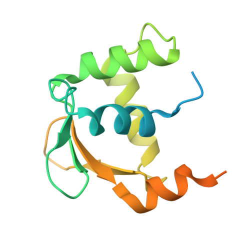
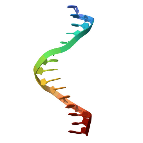
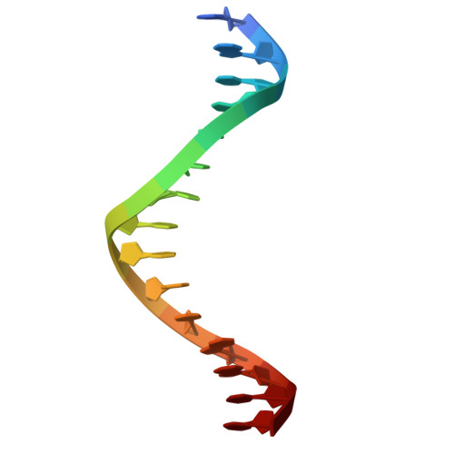
Structure and cooperative formation of a FLI1 filament on contiguous GGAA DNA sites.

Hou, C.Tsodikov, O.V.

(2025) Nucleic Acids Res 53

  • DOI: https://doi.org/10.1093/nar/gkaf205
  • Primary Citation of Related Structures:  
    9CP6, 9MWY, 9MX8, 9MX9, 9MXA

  • PubMed Abstract: 

    Ewing sarcoma, a pediatric cancer of bone and soft tissue, is driven in most cases by an abnormal oncogenic fusion of the N-terminal region of EWS with the C-terminal region of FLI1 (EWS-FLI1). The FLI1 region contains a conserved DNA-binding domain (DBD) essential for the oncogenesis. Binding of EWS-FLI1 to microsatellites composed of contiguous GGAA sites, shown previously to be critical for the oncogenic program of this fusion, is not well understood. In this study, we demonstrate that the FLI1 DBD binds cooperatively to contiguous GGAA sites, thereby forming a nucleoprotein filament. A series of crystal structures of two, three, and four FLI1 DBD proteins in complexes with DNA oligomers containing two, three, and four contiguous GGAA sites, respectively, reveal the structure of this filament and the basis for its cooperative formation. We expect this mechanistic insight to be an important milestone in our understanding of the oncogenic function of EWS-FLI1 and exploiting it as a drug target.


  • Organizational Affiliation
    • Department of Pharmaceutical Sciences, College of Pharmacy, University of Kentucky, Lexington, KY 40536, United States.

Macromolecules

Find similar proteins by:  (by identity cutoff)  |  3D Structure
Entity ID: 1
MoleculeChains Sequence LengthOrganismDetailsImage
Friend leukemia integration 1 transcription factorA,
B [auth D]
145Homo sapiensMutation(s): 1 
Gene Names: FLI1
UniProt & NIH Common Fund Data Resources
Find proteins for Q01543 (Homo sapiens)
Explore Q01543 
Go to UniProtKB:  Q01543
PHAROS:  Q01543
GTEx:  ENSG00000151702 
Entity Groups  
Sequence Clusters30% Identity50% Identity70% Identity90% Identity95% Identity100% Identity
UniProt GroupQ01543
Sequence Annotations
Expand
  • Reference Sequence

Find similar nucleic acids by:  Sequence   |   3D Structure  

Entity ID: 2
MoleculeChains LengthOrganismImage
DNA (5'-D(*GP*AP*CP*CP*GP*GP*AP*AP*GP*GP*AP*AP*GP*TP*G)-3')C [auth E]15synthetic construct
Sequence Annotations
Expand
  • Reference Sequence

Find similar nucleic acids by:  Sequence   |   3D Structure  

Entity ID: 3
MoleculeChains LengthOrganismImage
DNA (5'-D(*CP*AP*CP*TP*TP*CP*CP*TP*TP*CP*CP*GP*GP*TP*C)-3')D [auth F]15synthetic construct
Sequence Annotations
Expand
  • Reference Sequence
Experimental Data & Validation

Experimental Data

  • Method: X-RAY DIFFRACTION
  • Resolution: 2.55 Å
  • R-Value Free:  0.250 (Depositor), 0.255 (DCC) 
  • R-Value Work:  0.206 (Depositor), 0.213 (DCC) 
  • R-Value Observed: 0.209 (Depositor) 
Space Group: I 41 2 2
Unit Cell:
Length ( Å )Angle ( ˚ )
a = 119α = 90
b = 119β = 90
c = 95.316γ = 90
Software Package:
Software NamePurpose
REFMACrefinement
HKL-2000data reduction
HKL-2000data scaling
PHASERphasing

Structure Validation

View Full Validation Report


Entry History & Funding Information

Deposition Data


Funding OrganizationLocationGrant Number
National Institutes of Health/National Cancer Institute (NIH/NCI)United StatesCA243529

Revision History  (Full details and data files)

  • Version 1.0: 2025-03-12
    Type: Initial release
  • Version 1.1: 2025-04-23
    Changes: Database references