


The X-ray diffraction structure of photosensitizer protein PSP2
Zhao, Y.M., Qi, C.To be published.
Experimental Data Snapshot
Starting Model: experimental
View more details
wwPDB Validation 3D Report Full Report

Entity ID: 1 | |||||
|---|---|---|---|---|---|
| Molecule | Chains | Sequence Length | Organism | Details | Image |
| PSP2 | 225 | Aequorea victoria | Mutation(s): 0 | 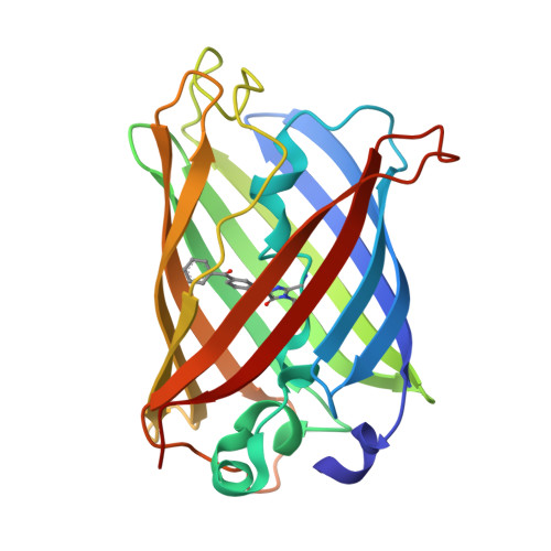 | |
UniProt | |||||
Entity Groups | |||||
| Sequence Clusters | 30% Identity50% Identity70% Identity90% Identity95% Identity100% Identity | ||||
| UniProt Group | P42212 | ||||
Sequence AnnotationsExpand | |||||
Reference Sequence | |||||
| Modified Residues 1 Unique | |||||
|---|---|---|---|---|---|
| ID | Chains | Type | Formula | 2D Diagram | Parent |
| BF6 Query on BF6 | A | L-PEPTIDE LINKING | C20 H17 N3 O4 |  | GLY, TYR, GLY |
| Length ( Å ) | Angle ( ˚ ) |
|---|---|
| a = 51.04 | α = 90 |
| b = 51.04 | β = 90 |
| c = 179.28 | γ = 90 |
| Software Name | Purpose |
|---|---|
| PHENIX | refinement |
| iMOSFLM | data reduction |
| Aimless | data scaling |
| PHASER | phasing |
| Funding Organization | Location | Grant Number |
|---|---|---|
| Ministry of Science and Technology (MoST, China) | China | 2019YFA0904101 |


