9LPU | pdb_00009lpu

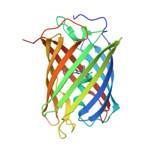
Crystal structure of tKeima at pH 4.0


Experimental Data Snapshot

  • Method: X-RAY DIFFRACTION
  • Resolution: 2.20 Å
  • R-Value Free: 
    0.275 (Depositor), 0.276 (DCC) 
  • R-Value Work: 
    0.231 (Depositor), 0.234 (DCC) 
  • R-Value Observed: 
    0.234 (Depositor) 

Starting Model: experimental
View more details

wwPDB Validation 3D Report Full Report

Validation slider image for 9LPU

This is version 2.0 of the entry. See complete history

Literature

pH-Induced Conformational Change of the Chromophore of the Large Stokes Shift Fluorescent Protein tKeima.

Xu, Y.Seo, Y.G.Kim, I.J.Nam, K.H.

(2025) Molecules 30

  • DOI: https://doi.org/10.3390/molecules30071623
  • Primary Citation Related Structures: 
    9LPU

  • PubMed Abstract: 

    Fluorescent proteins (FPs) are widely used as optical probes in molecular and cell biology. tKeima is a tetrameric, large Stokes shift red fluorescent protein and the ancestral protein of mt-Keima, which is widely applied as a pH-sensitive fluorescent probe. While the pH sensitivity of mt-Keima is well characterized, the pH-dependent properties of the ancestral tKeima have not been comprehensively elucidated. To obtain a better understanding of the effects of pH on tKeima, its fluorescent emission intensity at various pH levels was measured, and its crystal structure at pH 4.0 was determined at a resolution of 2.2 Å. The fluorescence emission intensity of tKeima at pH 4.0 decreased by approximately 65% compared with its peak emission at pH 10.0. The crystal structure of tKeima at pH 4.0 revealed both cis and trans conformations of the chromophore, in contrast to previously determined structures at pH 8.0, which showed only the cis conformation. This indicates that pH induces a conformational change of the chromophore in tKeima. Both the cis and trans conformations in tKeima were stabilized by hydrogen bonds with neighboring residues. A comparison of tKeima at pH 4.0 with tKeima at basic pH, as well as with mKeima, highlights its unique structural properties. These results provide a deeper understanding of the structural basis for the pH-induced fluorescence emission changes in the Keima family.


  • Organizational Affiliation
    • Department of Bioengineering, College of Life Science, Dalian Minzu University, Dalian 116600, China.

Macromolecule Content 

  • Total Structure Weight: 50.68 kDa 
  • Atom Count: 3,922 
  • Modeled Residue Count: 444 
  • Deposited Residue Count: 444 
  • Unique protein chains: 1

Macromolecules

Find similar proteins by:|  3D Structure
Entity ID: 1
MoleculeChains  Sequence LengthOrganismDetailsImage
Large stokes shift fluorescent protein
A, B
222Montipora sp. 20Mutation(s): 1 
Gene Names: t-keima
UniProt
Find proteins for Q1JV72 (Montipora sp. 20)
Explore Q1JV72 
Go to UniProtKB:  Q1JV72
Entity Groups
Sequence Clusters30% Identity50% Identity70% Identity90% Identity95% Identity100% Identity
UniProt GroupQ1JV72
Sequence Annotations
Expand
Reference Sequence

Small Molecules

Modified Residues  1 Unique
IDChains TypeFormula2D DiagramParent
CRQ
Query on CRQ
A, B
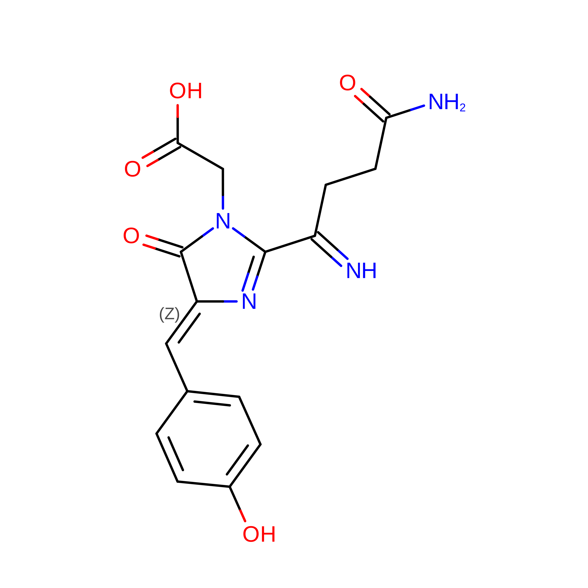
L-PEPTIDE LINKINGC16 H16 N4 O5GLN, TYR, GLY

Experimental Data & Validation

Experimental Data

  • Method: X-RAY DIFFRACTION
  • Resolution: 2.20 Å
  • R-Value Free:  0.275 (Depositor), 0.276 (DCC) 
  • R-Value Work:  0.231 (Depositor), 0.234 (DCC) 
  • R-Value Observed: 0.234 (Depositor) 
Space Group: P 21 21 2
Unit Cell:
Length ( Å )Angle ( ˚ )
a = 70.005α = 90
b = 85.773β = 90
c = 109.759γ = 90
Software Package:
Software NamePurpose
PHENIXrefinement
HKL-2000data scaling
HKL-2000data reduction
PHENIXphasing

Structure Validation

View Full Validation Report


Entry History 

& Funding Information

Deposition Data

  • Released Date: 2025-04-23 
  • Deposition Author(s): Nam, K.H.

Funding OrganizationLocationGrant Number
National Research Foundation (NRF, Korea)Korea, Republic OfNRF-2021R1I1A1A01050838

Revision History  (Full details and data files)

  • Version 1.0: 2025-04-23
    Type: Initial release
  • Version 1.1: 2025-05-14
    Changes: Database references
  • Version 2.0: 2026-03-18
    Changes: Polymer sequence