9JU0 | pdb_00009ju0

C-terminally truncated dextran dextrinase bound with acarbose


Experimental Data Snapshot

  • Method: X-RAY DIFFRACTION
  • Resolution: 2.50 Å
  • R-Value Free: 
    0.211 (Depositor), 0.214 (DCC) 
  • R-Value Work: 
    0.182 (Depositor), 0.186 (DCC) 

Starting Model: experimental
View more details

wwPDB Validation   3D Report Full Report


This is version 1.0 of the entry. See complete history


Literature

Structural basis of transglucosylation in dextran dextrinase, a homolog of anomer-inverting GH15 glucoside hydrolases.

Tagami, T.Saburi, W.Sadahiro, J.Kumagai, Y.Lang, W.Matsugaki, N.Okuyama, M.Mori, H.Kimura, A.

(2025) J Biological Chem 301: 110541-110541

  • DOI: https://doi.org/10.1016/j.jbc.2025.110541
  • Primary Citation of Related Structures:  
    9JU0

  • PubMed Abstract: 

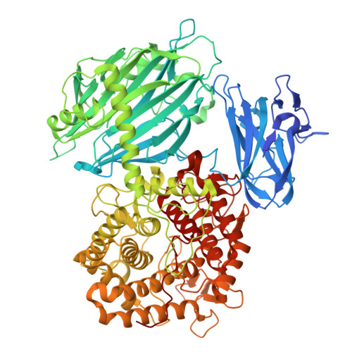
    Bacterial exopolysaccharide, dextran, primarily composed of α-(1→6)-linked d-glucosyl residues, is synthesized from α-(1→4)-glucan dextrin or sucrose through successive anomer-retaining transglucosylation reactions by dextran dextrinase (DDase) or dextransucrase, respectively. Although the structure-function relationship of dextransucrase has been extensively studied, that of DDase remains largely unknown. Herein, we revealed the Gluconobacter oxydans DDase structural basis through biochemical and structural analyses. The DDase comprises 1284 residues, with its N-terminal 902 residues being functionally essential. Crystal structure analysis of the minimal active DDase (Δ382C) complex with the pseudo-maltotetraose inhibitor, acarbose, revealed its homodimeric structure. A Δ382C protomer contains two β-sandwich domains, N1 and N2, and an (α/α) 6 -barrel domain A. Surprisingly, domains N2, A, and the helix-loop-helix connecting them structurally resemble those of bacterial anomer-inverting glucohydrolases in glycoside hydrolase family 15 (GH15). Domain N1 primarily forms intra- and inter-subunit domain interfaces. The DDase acarbose-binding residues in subsite -1 are conserved with GH15 glucohydrolases. The DDase Glu671 and Glu858 are positioned similarly to the GH15 glucohydrolase general acid and base catalysts, respectively. However, Glu858 is approximately 1.2 to 1.6 Å closer to the acarbose equivalent anomeric carbon, facilitating its role as a nucleophilic catalyst in the double displacement mechanism. The catalytic residue functions were biochemically confirmed using mutant enzymes. Spatial position of Glu858 is arranged by the local structure of the α11→α12 loop and subunit interactions involving domain N1. Enzymes classified in the same GH family catalyze reactions with different mechanisms, anomer-inverting or -retaining, due to differences in their catalytic residue spatial arrangement.


  • Organizational Affiliation
    • Research Faculty of Agriculture, Hokkaido University, Sapporo, Japan. Electronic address: tagami@agr.hokudai.ac.jp.

Macromolecules
Find similar proteins by:  (by identity cutoff)  |  3D Structure
Entity ID: 1
MoleculeChains Sequence LengthOrganismDetailsImage
Dextran dextrinaseA [auth AAA],
B [auth BBB],
C [auth CCC],
D [auth DDD]
923Gluconobacter oxydansMutation(s): 0 
Gene Names: EDC20_1461
EC: 2.4.1.2
UniProt
Find proteins for A0A0M5MSC0 (Gluconobacter oxydans)
Explore A0A0M5MSC0 
Go to UniProtKB:  A0A0M5MSC0
Entity Groups  
Sequence Clusters30% Identity50% Identity70% Identity90% Identity95% Identity100% Identity
UniProt GroupA0A0M5MSC0
Sequence Annotations
Expand
  • Reference Sequence
Oligosaccharides

Help

Entity ID: 2
MoleculeChains Length2D Diagram Glycosylation3D Interactions
4,6-dideoxy-4-{[(1S,4R,5S,6S)-4,5,6-trihydroxy-3-(hydroxymethyl)cyclohex-2-en-1-yl]amino}-alpha-D-glucopyranose-(1-4)-alpha-D-glucopyranose-(1-4)-alpha-D-glucopyranoseE [auth AaA],
F [auth BaB],
G [auth CaC],
H [auth DaD]
3N/AN/A
Glycosylation Resources
GlyTouCan:  G66431MI
GlyCosmos:  G66431MI
Small Molecules
Ligands 2 Unique
IDChains Name / Formula / InChI Key2D Diagram3D Interactions
CIT
Query on CIT

Download Ideal Coordinates CCD File 
IA [auth DDD],
Z [auth BBB]
CITRIC ACID
C6 H8 O7
KRKNYBCHXYNGOX-UHFFFAOYSA-N
EDO
Query on EDO

Download Ideal Coordinates CCD File 
AA [auth BBB]
BA [auth BBB]
CA [auth CCC]
DA [auth CCC]
EA [auth CCC]
AA [auth BBB],
BA [auth BBB],
CA [auth CCC],
DA [auth CCC],
EA [auth CCC],
FA [auth CCC],
GA [auth CCC],
HA [auth CCC],
I [auth AAA],
J [auth AAA],
JA [auth DDD],
K [auth AAA],
KA [auth DDD],
L [auth AAA],
M [auth AAA],
N [auth AAA],
O [auth AAA],
P [auth AAA],
Q [auth AAA],
R [auth AAA],
S [auth AAA],
T [auth AAA],
U [auth AAA],
V [auth AAA],
W [auth BBB],
X [auth BBB],
Y [auth BBB]
1,2-ETHANEDIOL
C2 H6 O2
LYCAIKOWRPUZTN-UHFFFAOYSA-N
Experimental Data & Validation

Experimental Data

  • Method: X-RAY DIFFRACTION
  • Resolution: 2.50 Å
  • R-Value Free:  0.211 (Depositor), 0.214 (DCC) 
  • R-Value Work:  0.182 (Depositor), 0.186 (DCC) 
Space Group: P 21 21 21
Unit Cell:
Length ( Å )Angle ( ˚ )
a = 118.259α = 90
b = 138.969β = 90
c = 264.462γ = 90
Software Package:
Software NamePurpose
REFMACrefinement
PHENIXrefinement
XDSdata reduction
XDSdata scaling
PHASERphasing

Structure Validation

View Full Validation Report


Entry History & Funding Information

Deposition Data


Funding OrganizationLocationGrant Number
Japan Society for the Promotion of Science (JSPS)Japan--

Revision History  (Full details and data files)

  • Version 1.0: 2025-09-03
    Type: Initial release