9JM1 | pdb_00009jm1

Crystal structure of de novo designed light-responsive oligomer C2-5 (LRO-C2-5)


Experimental Data Snapshot

  • Method: X-RAY DIFFRACTION
  • Resolution: 2.40 Å
  • R-Value Free: 
    0.334 (Depositor), 0.333 (DCC) 
  • R-Value Work: 
    0.299 (Depositor), 0.299 (DCC) 
  • R-Value Observed: 
    0.301 (Depositor) 

Starting Model: in silico
View more details

wwPDB Validation   3D Report Full Report


This is version 1.2 of the entry. See complete history


Literature

De novo design of light-responsive protein-protein interactions enables reversible formation of protein assemblies.

Yu, B.Liu, J.Cui, Z.Wang, C.Chen, P.Wang, C.Zhang, Y.Zhu, X.Zhang, Z.Li, S.Pan, J.Xie, M.Shen, H.Cao, L.

(2025) Nat Chem 17: 1910-1919

  • DOI: https://doi.org/10.1038/s41557-025-01929-2
  • Primary Citation of Related Structures:  
    9JM1, 9JM2, 9JM3, 9JM4, 9JM5, 9JM6, 9JM7, 9JM8

  • PubMed Abstract: 

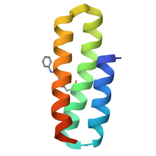
    Light-responsive proteins play an essential role in all domains of life by sensing and responding to environmental light signals. However, the de novo design of light-responsive proteins with precisely defined structures and reversible responsive behaviours is an unmet challenge. Here we describe a computational approach to design protein-protein interactions regulated by non-canonical amino acids, focusing on the light-responsive phenylalanine-4'-azobenzene (AzoF). Using this approach, we designed light-responsive cyclic homo-oligomers and heterodimers, which only assemble in AzoF's trans configuration and disassemble when AzoF photoisomerizes to the cis configuration. Biophysical characterization confirms the light-responsive assembly and disassembly of these complexes, and the crystal structures match the design models with atomic accuracy. We demonstrate the applicability of these light-responsive proteins in constructing light-responsive hydrogels and engineering synthetic ligand receptors to optocontrol cell signalling in mammalian cells. Our approach opens avenues for designing environmentally responsive protein structures and broadens the toolkit for optogenetics and optochemistry.


  • Organizational Affiliation
    • School of Life Sciences, Westlake University, Hangzhou, China.

Macromolecules
Find similar proteins by:  (by identity cutoff)  |  3D Structure
Entity ID: 1
MoleculeChains Sequence LengthOrganismDetailsImage
light-responsive oligomer C2-5
A, B, C, D, E
73Escherichia coliMutation(s): 0 
Entity Groups  
Sequence Clusters30% Identity50% Identity70% Identity90% Identity95% Identity100% Identity
Sequence Annotations
Expand
  • Reference Sequence
Small Molecules
Modified Residues  1 Unique
IDChains TypeFormula2D DiagramParent
OZW
Query on OZW
A, B, C, D, E
L-PEPTIDE LINKINGC15 H17 N3 O2PHE
Experimental Data & Validation

Experimental Data

  • Method: X-RAY DIFFRACTION
  • Resolution: 2.40 Å
  • R-Value Free:  0.334 (Depositor), 0.333 (DCC) 
  • R-Value Work:  0.299 (Depositor), 0.299 (DCC) 
  • R-Value Observed: 0.301 (Depositor) 
Space Group: P 31 2 1
Unit Cell:
Length ( Å )Angle ( ˚ )
a = 75.748α = 90
b = 75.748β = 90
c = 130.03γ = 120
Software Package:
Software NamePurpose
PHENIXrefinement
PHENIXphasing
CrysalisProdata reduction
CrysalisProdata scaling

Structure Validation

View Full Validation Report


Entry History & Funding Information

Deposition Data

  • Released Date: 2025-08-27 
  • Deposition Author(s): Yu, B., Cao, L.

Funding OrganizationLocationGrant Number
Ministry of Science and Technology (MoST, China)China2022YFA1303700

Revision History  (Full details and data files)

  • Version 1.0: 2025-08-27
    Type: Initial release
  • Version 1.1: 2025-09-10
    Changes: Database references
  • Version 1.2: 2025-12-17
    Changes: Database references