9J22 | pdb_00009j22

structure of human urea transport protein slc14A1 with urea


Experimental Data Snapshot

  • Method: ELECTRON MICROSCOPY
  • Resolution: 2.75 Å
  • Aggregation State: PARTICLE 
  • Reconstruction Method: SINGLE PARTICLE 

wwPDB Validation 3D Report Full Report

Validation slider image for 9J22

This is version 1.0 of the entry. See complete history

Literature

Structural basis of the bifunctionality of Marinobacter salinexigens ZYF650 T glucosylglycerol phosphorylase in glucosylglycerol catabolism.

Lu, D.Zhang, K.Cheng, C.Wu, D.Yin, L.Luo, Q.Shi, M.Ma, H.Lu, X.

(2025) J Biological Chem 301: 108127-108127

  • DOI: https://doi.org/10.1016/j.jbc.2024.108127
  • Primary Citation Related Structures: 
    9J1U, 9J22, 9J24, 9J25

  • PubMed Abstract: 

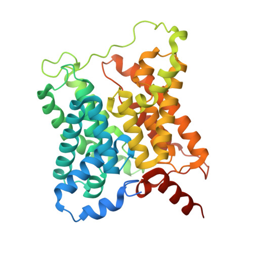
    2-O-α-Glucosylglycerol (GG) is a natural heteroside synthesized by many cyanobacteria and a few heterotrophic bacteria under salt stress conditions. Bacteria produce GG in response to stimuli and degrade it once the stimulus diminishes. Heterotrophic bacteria utilize GG phosphorylase (GGP), a member of the GH13_18 family, via a two-step process consisting of phosphorolysis and hydrolysis for GG catabolism. However, the precise mechanism by which GGP degrades GG remains elusive. We determined the 3D structure of a recently identified GGP (MsGGP) of the deep-sea bacterium Marinobacter salinexigens ZYF650 T , in complex with glucose and glycerol, α-d-glucose-1-phosphate (αGlc1-P), and orthophosphate (inorganic phosphate) at resolutions of 2.5, 2.7, and 2.7 Å, respectively. Notably, the first αGlc1-P complex structure in the GH13_18 family, the complex of MsGGP and αGlc1-P, validates that GGP catalyzes GG decomposition through consecutive phosphorolysis and hydrolysis. In addition, the structure reveals the mechanism of high stereoselectivity on αGlc1-P. Glu231 and Asp190 were identified as the catalytic residues. Interestingly, these structures closely resemble each other, indicating minimal conformational changes upon binding end-product glucose and glycerol, or the intermediate αGlc1-P. The structures also indicate that the substrates may follow a specific trajectory and a precise order toward the active center in close proximity and in a geometrically favorable orientation for catalysis in a double displacement mechanism.


  • Organizational Affiliation
    • School of Chemical Engineering, Marine and Life Sciences, Dalian University of Technology, Panjin, China; Key Laboratory of Biofuels, Qingdao Institute of Bioenergy and Bioprocess Technology, Chinese Academy of Sciences, Qingdao, China.

Macromolecule Content 

  • Total Structure Weight: 131.68 kDa 
  • Atom Count: 8,238 
  • Modeled Residue Count: 1,041 
  • Deposited Residue Count: 1,167 
  • Unique protein chains: 1

Macromolecules

Find similar proteins by:|  3D Structure
Entity ID: 1
MoleculeChains  Sequence LengthOrganismDetailsImage
Urea transporter 1
A, B, C
389Homo sapiensMutation(s): 0 
Gene Names: SLC14A1HUT11JKRACH1UT1UTE
Membrane Entity: Yes 
UniProt & NIH Common Fund Data Resources
Find proteins for Q13336 (Homo sapiens)
Explore Q13336 
Go to UniProtKB:  Q13336
PHAROS:  Q13336
GTEx:  ENSG00000141469 
Entity Groups
Sequence Clusters30% Identity50% Identity70% Identity90% Identity95% Identity100% Identity
UniProt GroupQ13336
Sequence Annotations
Expand
Reference Sequence

Small Molecules

Ligands 2 Unique
IDChains Name / Formula / InChI Key2D Diagram3D Interactions
PLM
(Subject of Investigation/LOI)

Query on PLM


Download:Ideal Coordinates CCD File
BA [auth C]
CA [auth C]
DA [auth C]
I [auth A]
J [auth A]
BA [auth C],
CA [auth C],
DA [auth C],
I [auth A],
J [auth A],
K [auth A],
L [auth A],
M [auth A],
N [auth A],
T [auth B],
U [auth B],
V [auth B]
PALMITIC ACID
C16 H32 O2
IPCSVZSSVZVIGE-UHFFFAOYSA-N
URE
(Subject of Investigation/LOI)

Query on URE


Download:Ideal Coordinates CCD File
AA [auth C]
D [auth A]
E [auth A]
F [auth A]
G [auth A]
AA [auth C],
D [auth A],
E [auth A],
F [auth A],
G [auth A],
H [auth A],
O [auth B],
P [auth B],
Q [auth B],
R [auth B],
S [auth B],
W [auth C],
X [auth C],
Y [auth C],
Z [auth C]
UREA
C H4 N2 O
XSQUKJJJFZCRTK-UHFFFAOYSA-N

Experimental Data & Validation

Experimental Data

  • Method: ELECTRON MICROSCOPY
  • Resolution: 2.75 Å
  • Aggregation State: PARTICLE 
  • Reconstruction Method: SINGLE PARTICLE 
EM Software:
TaskSoftware PackageVersion
MODEL REFINEMENTPHENIX1.21rc1_5127:

Structure Validation

View Full Validation Report


Entry History 

& Funding Information

Deposition Data


Funding OrganizationLocationGrant Number
Other governmentChinaC10120180025

Revision History  (Full details and data files)

  • Version 1.0: 2025-09-03
    Type: Initial release