9ITI | pdb_00009iti

Nav1.7 with mutations that eliminate beta1 binding


Experimental Data Snapshot

  • Method: ELECTRON MICROSCOPY
  • Resolution: 2.92 Å
  • Aggregation State: PARTICLE 
  • Reconstruction Method: SINGLE PARTICLE 

wwPDB Validation   3D Report Full Report


This is version 1.0 of the entry. See complete history


Literature

Critical role of extracellular loops in differential modulations of TTX-sensitive and TTX-resistant Na v channels.

Wu, T.Yang, X.Jin, X.Yan, N.Li, Z.

(2025) Proc Natl Acad Sci U S A 122: e2510355122-e2510355122

  • DOI: https://doi.org/10.1073/pnas.2510355122
  • Primary Citation of Related Structures:  
    9ITH, 9ITI

  • PubMed Abstract: 

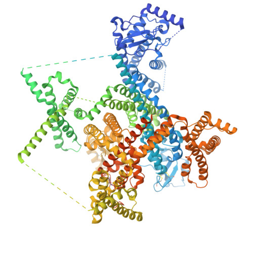
    The cardiac voltage-gated sodium channel Na v 1.5 is resistant to tetrodotoxin (TTXr). Here, we report a cryo-electron microscopy (cryo-EM) structure of wild-type human Na v 1.5, coexpressed with the β1 auxiliary subunit and treated with high-concentration TTX, at 3.4 Å resolution. Structural comparison reveals the molecular determinants for the distinct responses to TTX as well as β subunits between TTXr and TTX-sensitive (TTXs) Na v channels. A conserved cation-π interaction between the guanidinium group of TTX and Tyr or Phe on the P2 I helix in TTXs Na v channels is lost in all TTXr subtypes owing to the replacement by Cys/Ser at the corresponding locus, explaining their differential TTX sensitivities. The β1 subunit is invisible in the EM map. Comparison of Na v 1.5 with Na v 1.7 and Na v 1.8, which are, respectively, TTXs and TTXr, identifies four sites on the extracellular loops (ECLs) that may account for their different β1-binding abilities. When the corresponding residues in TTXs Na v 1.7 are replaced with those from Na v 1.5, the modulatory effects of β1 on channel activation and inactivation are diminished. Consistently, β1 is absent in the 3D EM reconstruction of this Na v 1.7 mutant. Together with our previous structure-guided discovery that TTXr channels lack a Cys on the ECL II for disulfide bond formation with β2 or β4, the structure-function relationship studies underscore the importance of the ECLs in the mechanistic distinctions between TTXs and TTXr Na v channels. The ECLs may be further explored for the development of subtype-specific drugs.


  • Organizational Affiliation
    • State Key Laboratory of Membrane Biology, Beijing Advanced Innovation Center for Structural Biology, Tsinghua-Peking Joint Center for Life Sciences, School of Life Sciences, Tsinghua University, Beijing 100084, China.

Macromolecules
Find similar proteins by:  (by identity cutoff)  |  3D Structure
Entity ID: 1
MoleculeChains Sequence LengthOrganismDetailsImage
Sodium channel protein type 9 subunit alpha2,030Homo sapiensMutation(s): 11 
Gene Names: SCN9ANENA
Membrane Entity: Yes 
UniProt & NIH Common Fund Data Resources
Find proteins for Q15858 (Homo sapiens)
Explore Q15858 
Go to UniProtKB:  Q15858
PHAROS:  Q15858
GTEx:  ENSG00000169432 
Entity Groups  
Sequence Clusters30% Identity50% Identity70% Identity90% Identity95% Identity100% Identity
UniProt GroupQ15858
Glycosylation
Glycosylation Sites: 4Go to GlyGen: Q15858-1
Sequence Annotations
Expand
  • Reference Sequence
Oligosaccharides

Help

Entity ID: 2
MoleculeChains Length2D Diagram Glycosylation3D Interactions
2-acetamido-2-deoxy-beta-D-glucopyranose-(1-4)-2-acetamido-2-deoxy-beta-D-glucopyranose
B, C
2N-Glycosylation
Glycosylation Resources
GlyTouCan:  G42666HT
GlyCosmos:  G42666HT
GlyGen:  G42666HT
Small Molecules
Ligands 8 Unique
IDChains Name / Formula / InChI Key2D Diagram3D Interactions
P5S
Query on P5S

Download Ideal Coordinates CCD File 
F [auth A],
P [auth A]
O-[(R)-{[(2R)-2,3-bis(octadecanoyloxy)propyl]oxy}(hydroxy)phosphoryl]-L-serine
C42 H82 N O10 P
TZCPCKNHXULUIY-RGULYWFUSA-N
PCW
Query on PCW

Download Ideal Coordinates CCD File 
L [auth A],
O [auth A],
Q [auth A],
W [auth A],
X [auth A]
1,2-DIOLEOYL-SN-GLYCERO-3-PHOSPHOCHOLINE
C44 H85 N O8 P
SNKAWJBJQDLSFF-NVKMUCNASA-O
9Z9
Query on 9Z9

Download Ideal Coordinates CCD File 
AA [auth A](3beta,14beta,17beta,25R)-3-[4-methoxy-3-(methoxymethyl)butoxy]spirost-5-en
C34 H56 O5
CEEBZAXXSRFQIC-GZSGZGDASA-N
LPE
Query on LPE

Download Ideal Coordinates CCD File 
H [auth A]
I [auth A]
K [auth A]
M [auth A]
N [auth A]
H [auth A],
I [auth A],
K [auth A],
M [auth A],
N [auth A],
R [auth A],
S [auth A],
T [auth A],
U [auth A],
V [auth A]
1-O-OCTADECYL-SN-GLYCERO-3-PHOSPHOCHOLINE
C26 H57 N O6 P
XKBJVQHMEXMFDZ-AREMUKBSSA-O
Y01
Query on Y01

Download Ideal Coordinates CCD File 
BA [auth A],
CA [auth A],
Y [auth A],
Z [auth A]
CHOLESTEROL HEMISUCCINATE
C31 H50 O4
WLNARFZDISHUGS-MIXBDBMTSA-N
1PW
Query on 1PW

Download Ideal Coordinates CCD File 
J [auth A](2S,3R,4E)-2-(acetylamino)-3-hydroxyoctadec-4-en-1-yl dihydrogen phosphate
C20 H40 N O6 P
HWPZKJVGDYNEAW-QUDYQQOWSA-N
NAG
Query on NAG

Download Ideal Coordinates CCD File 
D [auth A],
E [auth A]
2-acetamido-2-deoxy-beta-D-glucopyranose
C8 H15 N O6
OVRNDRQMDRJTHS-FMDGEEDCSA-N
NA
Query on NA

Download Ideal Coordinates CCD File 
G [auth A]SODIUM ION
Na
FKNQFGJONOIPTF-UHFFFAOYSA-N
Experimental Data & Validation

Experimental Data

  • Method: ELECTRON MICROSCOPY
  • Resolution: 2.92 Å
  • Aggregation State: PARTICLE 
  • Reconstruction Method: SINGLE PARTICLE 
EM Software:
TaskSoftware PackageVersion
MODEL REFINEMENTPHENIX1.18_3855:

Structure Validation

View Full Validation Report


Entry History & Funding Information

Deposition Data


Funding OrganizationLocationGrant Number
National Natural Science Foundation of China (NSFC)China32330052

Revision History  (Full details and data files)

  • Version 1.0: 2025-08-20
    Type: Initial release