9IT2 | pdb_00009it2

Cryo-EM structure of urease from Ureaplasma parvum


Experimental Data Snapshot

  • Method: ELECTRON MICROSCOPY
  • Resolution: 2.03 Å
  • Aggregation State: PARTICLE 
  • Reconstruction Method: SINGLE PARTICLE 

Starting Model: in silico
View more details

wwPDB Validation   3D Report Full Report


This is version 1.0 of the entry. See complete history


Literature

Structural Analysis and Molecular Dynamics Simulations of Urease From Ureaplasma parvum.

Wu, H.N.Fujita, J.Nakura, Y.Inoue, M.Suzuki, K.Ekimoto, T.Yin, B.Fukuda, Y.Harada, K.Inoue, T.Ikeguchi, M.Namba, K.Yanagihara, I.

(2025) J Mol Biology 437: 169368-169368

  • DOI: https://doi.org/10.1016/j.jmb.2025.169368
  • Primary Citation of Related Structures:  
    9IT2

  • PubMed Abstract: 

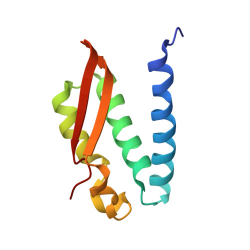
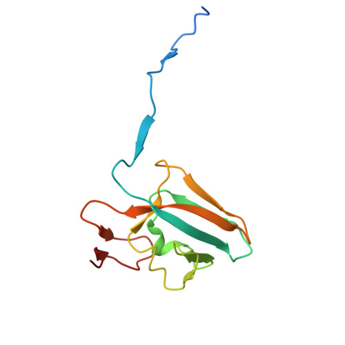
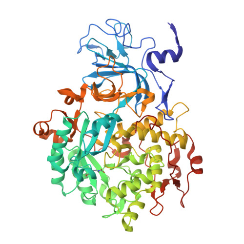
    Ureaplasma is one of the smallest pathogenic bacteria, generating approximately 95% of its adenosine triphosphate (ATP) solely through urease. Studies on Ureaplasma parvum, a species of Ureaplasma, have confirmed that adding urease inhibitors inhibits bacterial growth. The K m and V max of the urease-mediated reaction were estimated to be 4.3 ± 0.2 mM and 3,333.3 ± 38.0 μmol NH 3 /min/mg protein, respectively. The cryo-electron microscopy (cryo-EM) structure of Ureaplasma parvum urease (UPU) at a resolution of 2.03 Å reveals a trimer of heterotrimers comprising three proteins: UreA, UreB, and UreC. The active site is well conserved among the known ureases. However, the V max of UPU was higher than that of most known ureases, including those ureases derived from Sporosarcina pasteurii (SPU) and Klebsiella aerogenes (KAU) with identical oligomeric state. All-atom molecular dynamics simulations showed that the flap and UreB are more open in UPU than SPU and KAU. His-tagged wild-type recombinant UPU (WT-rUPU) revealed estimated K m and V max values of 4.1 ± 0.3 mM and 769.2 ± 7.4 µmol NH 3 /min/mg protein, respectively. Amino acid substitutions of recombinant UPUs within the flap region to SPU. Amongst the flap region variants, the V max of K331N variant was 48-fold lower than that of WT-rUPU. ICP-MS analysis reveals that one molecule of UPU, WT-rUPU, and K331N-rUPU contains 3.7, 0.8, and 0.1 Ni 2+ atoms, respectively, suggesting that a wide-open flap of urease may contribute to delivering nickel into the enzyme, resulting in a high V max . Ureaplasma evolved highly efficient UPU through a few amino acid substitutions in the disorganized loop of the mobile flap region.


  • Organizational Affiliation
    • Department of Developmental Medicine, Research Institute, Osaka Women's and Children's Hospital, Izumi City 594-1101 Osaka, Japan.

Macromolecules
Find similar proteins by:  (by identity cutoff)  |  3D Structure
Entity ID: 1
MoleculeChains Sequence LengthOrganismDetailsImage
Urease subunit gamma
A, D, G
101Ureaplasma parvum serovar 3 str. ATCC 700970Mutation(s): 0 
EC: 3.5.1.5
UniProt
Find proteins for P0C7K9 (Ureaplasma parvum serovar 3 (strain ATCC 700970))
Explore P0C7K9 
Go to UniProtKB:  P0C7K9
Entity Groups  
Sequence Clusters30% Identity50% Identity70% Identity90% Identity95% Identity100% Identity
UniProt GroupP0C7K9
Sequence Annotations
Expand
  • Reference Sequence
Find similar proteins by:  (by identity cutoff)  |  3D Structure
Entity ID: 2
MoleculeChains Sequence LengthOrganismDetailsImage
Urease subunit beta
B, E, H
124Ureaplasma parvum serovar 3 str. ATCC 700970Mutation(s): 0 
EC: 3.5.1.5
UniProt
Find proteins for P0C7K8 (Ureaplasma parvum serovar 3 (strain ATCC 700970))
Explore P0C7K8 
Go to UniProtKB:  P0C7K8
Entity Groups  
Sequence Clusters30% Identity50% Identity70% Identity90% Identity95% Identity100% Identity
UniProt GroupP0C7K8
Sequence Annotations
Expand
  • Reference Sequence
Find similar proteins by:  (by identity cutoff)  |  3D Structure
Entity ID: 3
MoleculeChains Sequence LengthOrganismDetailsImage
Urease subunit alpha
C, F, I
598Ureaplasma parvum serovar 3 str. ATCC 700970Mutation(s): 0 
EC: 3.5.1.5
UniProt
Find proteins for P0C7K7 (Ureaplasma parvum serovar 3 (strain ATCC 700970))
Explore P0C7K7 
Go to UniProtKB:  P0C7K7
Entity Groups  
Sequence Clusters30% Identity50% Identity70% Identity90% Identity95% Identity100% Identity
UniProt GroupP0C7K7
Sequence Annotations
Expand
  • Reference Sequence
Small Molecules
Ligands 2 Unique
IDChains Name / Formula / InChI Key2D Diagram3D Interactions
BME (Subject of Investigation/LOI)
Query on BME

Download Ideal Coordinates CCD File 
J [auth C],
M [auth F],
P [auth I]
BETA-MERCAPTOETHANOL
C2 H6 O S
DGVVWUTYPXICAM-UHFFFAOYSA-N
NI (Subject of Investigation/LOI)
Query on NI

Download Ideal Coordinates CCD File 
K [auth C]
L [auth C]
N [auth F]
O [auth F]
Q [auth I]
K [auth C],
L [auth C],
N [auth F],
O [auth F],
Q [auth I],
R [auth I]
NICKEL (II) ION
Ni
VEQPNABPJHWNSG-UHFFFAOYSA-N
Modified Residues  1 Unique
IDChains TypeFormula2D DiagramParent
KCX
Query on KCX
C, F, I
L-PEPTIDE LINKINGC7 H14 N2 O4LYS
Experimental Data & Validation

Experimental Data

  • Method: ELECTRON MICROSCOPY
  • Resolution: 2.03 Å
  • Aggregation State: PARTICLE 
  • Reconstruction Method: SINGLE PARTICLE 
EM Software:
TaskSoftware PackageVersion
RECONSTRUCTIONcryoSPARC4.2.1
MODEL REFINEMENTPHENIX1.19.1-4122

Structure Validation

View Full Validation Report


Entry History & Funding Information

Deposition Data


Funding OrganizationLocationGrant Number
Japan Agency for Medical Research and Development (AMED)JapanJP23fk0108677
Japan Agency for Medical Research and Development (AMED)JapanJP21am0101117 and JP22ama121003
Japan Agency for Medical Research and Development (AMED)JapanJP24ama121023
Japan Agency for Medical Research and Development (AMED)JapanJP17pc0101020
Japan Science and TechnologyJapanJPMJOP1861
The Research Foundation for Microbial Diseases, Osaka UniversityJapan--
JEOL YOKOGUSHI Research Alliance Laboratories of Osaka UniversityJapan--

Revision History  (Full details and data files)

  • Version 1.0: 2025-08-20
    Type: Initial release