9HVW | pdb_00009hvw

Respiratory Syncytial Virus Fusion protein in the postfusion conformation in complex with monoclonal antibody 131-2a Fab


Experimental Data Snapshot

  • Method: ELECTRON MICROSCOPY
  • Resolution: 3.10 Å
  • Aggregation State: PARTICLE 
  • Reconstruction Method: SINGLE PARTICLE 

wwPDB Validation   3D Report Full Report


This is version 1.2 of the entry. See complete history


Literature

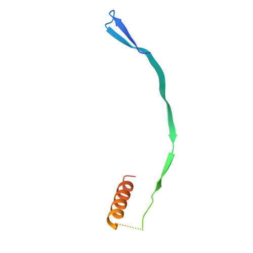
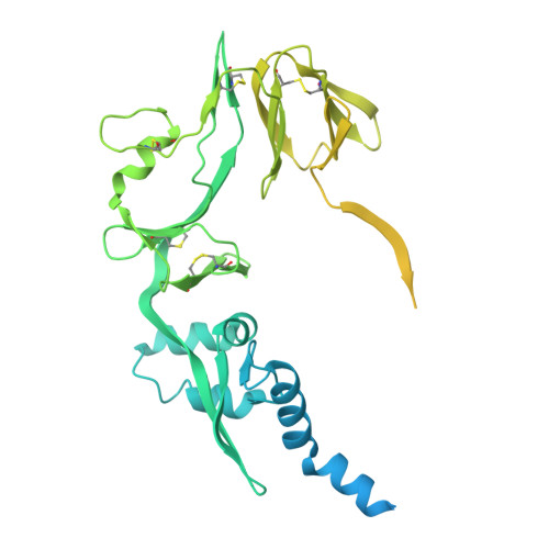
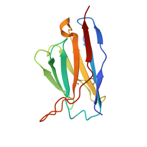
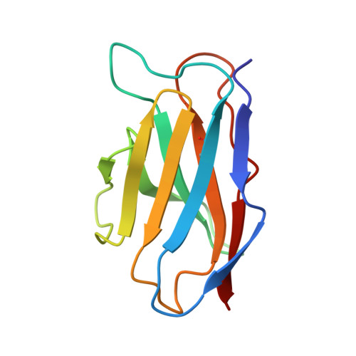
Structural Basis for Postfusion-Specific Binding to the Respiratory Syncytial Virus F Protein by the Canonical Antigenic Site I Antibody 131-2a.

Peng, W.Siborova, M.Wu, X.Du, W.Schulte, D.Pronker, M.F.de Haan, C.A.M.Snijder, J.

(2025) ACS Infect Dis 11: 2357-2366

  • DOI: https://doi.org/10.1021/acsinfecdis.5c00368
  • Primary Citation of Related Structures:  
    9HVW

  • PubMed Abstract: 

    The respiratory syncytial virus (RSV) fusion (F) protein is a major target of antiviral antibodies following natural infection or vaccination and is responsible for mediating fusion between the viral envelope and the host membrane. The fusion process is driven by a large-scale conformational change in F, switching irreversibly from the metastable prefusion state to the stable postfusion conformation. Previous research has identified six distinct antigenic sites in RSV-F, termed sites Ø, I, II, III, IV, and V. Of these, only antigenic site I is fully specific to the postfusion conformation of F. A monoclonal antibody 131-2a that specifically targets postfusion F has been widely used as a research tool to probe for postfusion F and to define antigenic site I in serological studies, yet its sequence and precise epitope have remained unknown. Here, we use mass spectrometry-based de novo sequencing of 131-2a to reverse engineer a recombinant product and study the epitope to define antigenic site I with molecular detail, revealing the structural basis for the antibody's specificity toward postfusion RSV-F.


  • Organizational Affiliation
    • Biomolecular Mass Spectrometry and Proteomics, Bijvoet Center for Biomolecular Research and Utrecht Institute of Pharmaceutical Sciences, Utrecht University, Padualaan 8, Utrecht 3584CH, the Netherlands.

Macromolecules
Find similar proteins by:  (by identity cutoff)  |  3D Structure
Entity ID: 1
MoleculeChains Sequence LengthOrganismDetailsImage
Fusion glycoprotein F0
A, B, C
80human respiratory syncytial virusMutation(s): 1 
UniProt
Find proteins for P03420 (Human respiratory syncytial virus A (strain A2))
Explore P03420 
Go to UniProtKB:  P03420
Entity Groups  
Sequence Clusters30% Identity50% Identity70% Identity90% Identity95% Identity100% Identity
UniProt GroupP03420
Sequence Annotations
Expand
  • Reference Sequence
Find similar proteins by:  (by identity cutoff)  |  3D Structure
Entity ID: 2
MoleculeChains Sequence LengthOrganismDetailsImage
Fusion glycoprotein F1,Probable N-acetylmuramidase
D, E, F
511human respiratory syncytial virusMutation(s): 0 
EC: 3.2.1.17
UniProt
Find proteins for A2RHZ5 (Lactococcus lactis subsp. cremoris (strain MG1363))
Explore A2RHZ5 
Go to UniProtKB:  A2RHZ5
Find proteins for P03420 (Human respiratory syncytial virus A (strain A2))
Explore P03420 
Go to UniProtKB:  P03420
Entity Groups  
Sequence Clusters30% Identity50% Identity70% Identity90% Identity95% Identity100% Identity
UniProt GroupsA2RHZ5P03420
Sequence Annotations
Expand
  • Reference Sequence
Find similar proteins by:  (by identity cutoff)  |  3D Structure
Entity ID: 3
MoleculeChains Sequence LengthOrganismDetailsImage
131-2a heavy chainG [auth H]120Mus musculusMutation(s): 0 
Entity Groups  
Sequence Clusters30% Identity50% Identity70% Identity90% Identity95% Identity100% Identity
Sequence Annotations
Expand
  • Reference Sequence
Find similar proteins by:  (by identity cutoff)  |  3D Structure
Entity ID: 4
MoleculeChains Sequence LengthOrganismDetailsImage
131-2a light chainH [auth L]111Mus musculusMutation(s): 0 
Entity Groups  
Sequence Clusters30% Identity50% Identity70% Identity90% Identity95% Identity100% Identity
Sequence Annotations
Expand
  • Reference Sequence
Experimental Data & Validation

Experimental Data

  • Method: ELECTRON MICROSCOPY
  • Resolution: 3.10 Å
  • Aggregation State: PARTICLE 
  • Reconstruction Method: SINGLE PARTICLE 

Structure Validation

View Full Validation Report


Entry History & Funding Information

Deposition Data

  • Released Date: 2025-02-26 
  • Deposition Author(s): Snijder, J.

Funding OrganizationLocationGrant Number
Netherlands Organisation for Scientific Research (NWO)Netherlands--
European Research Council (ERC)European Union--

Revision History  (Full details and data files)

  • Version 1.0: 2025-02-26
    Type: Initial release
  • Version 1.1: 2025-08-06
    Changes: Data collection, Database references
  • Version 1.2: 2025-08-20
    Changes: Data collection, Database references