9HQU | pdb_00009hqu

Salmonella enterica Lamassu LmuACB in nuclease sequestration state


Experimental Data Snapshot

  • Method: ELECTRON MICROSCOPY
  • Resolution: 3.21 Å
  • Aggregation State: 3D ARRAY 
  • Reconstruction Method: SINGLE PARTICLE 

wwPDB Validation   3D Report Full Report


This is version 1.3 of the entry. See complete history


Literature

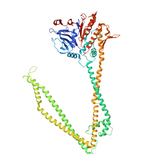
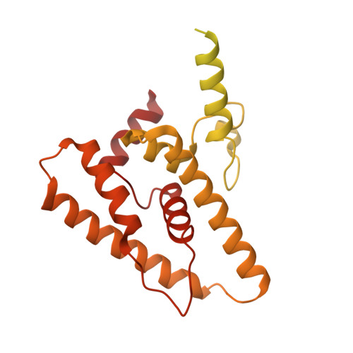
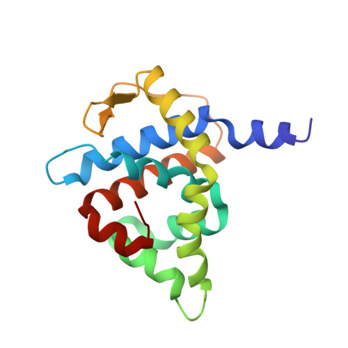
Structure and activation mechanism of a Lamassu phage and plasmid defense system.

Li, Y.Adams, D.W.Liu, H.W.Shaw, S.J.Uchikawa, E.Jaskolska, M.Stutzmann, S.Righi, L.Szczelkun, M.D.Blokesch, M.Gruber, S.

(2025) Nat Struct Mol Biol 32: 2503-2516

  • DOI: https://doi.org/10.1038/s41594-025-01677-4
  • Primary Citation of Related Structures:  
    9HQU, 9HQX, 9HR5

  • PubMed Abstract: 

    Lamassu is a diverse family of defense systems that protect bacteria, including seventh-pandemic strains of Vibrio cholerae, against both plasmids and phage infection. During phage infection, Lamassu targets essential cellular processes, thereby halting phage propagation by terminating the infected host. The mechanisms by which Lamassu effectors are activated when needed and otherwise suppressed are unknown. Here we present structures of a Lamassu defense system from Salmonella enterica. We show that an oligomerization domain of the nuclease effector subunit, LmuA, is sequestered by two tightly folded SMC-like LmuB protomers and LmuC. Upon activation, liberated LmuA assembles into homotetramers, in which two of four nuclease domains are brought into proximity to create an active site capable of cleaving DNA. We propose that tetramer formation is likely a one-way switch that establishes a threshold to limit potential spontaneous activation and cell death. Our findings reveal a mechanism of cellular defense, involving liberation and oligomerization of immune effectors, and shed light on how Lamassu systems balance potent immune responses with self-preservation.


  • Organizational Affiliation
    • Department of Fundamental Microbiology (DMF), Faculty of Biology and Medicine (FBM), University of Lausanne (UNIL), Lausanne, Switzerland.

Macromolecules
Find similar proteins by:  (by identity cutoff)  |  3D Structure
Entity ID: 1
MoleculeChains Sequence LengthOrganismDetailsImage
DUF3732 domain-containing protein
A, B
660Salmonella enterica subsp. enterica serovar TennesseeMutation(s): 0 
Gene Names: AHW86_02670B1277_19175CC786_01560DKV27_15540E0563_21155FG623_013450GB216_17200
UniProt
Find proteins for A0A3G3DSI6 (Salmonella enterica subsp. enterica serovar Tennessee)
Explore A0A3G3DSI6 
Go to UniProtKB:  A0A3G3DSI6
Entity Groups  
Sequence Clusters30% Identity50% Identity70% Identity90% Identity95% Identity100% Identity
UniProt GroupA0A3G3DSI6
Sequence Annotations
Expand
  • Reference Sequence
Find similar proteins by:  (by identity cutoff)  |  3D Structure
Entity ID: 2
MoleculeChains Sequence LengthOrganismDetailsImage
ABC-three component systems C-terminal domain-containing protein403Salmonella enterica subsp. enterica serovar TennesseeMutation(s): 0 
Gene Names: AHW86_02680B1277_19165CC786_01570DKV27_15550E0563_21145FG623_013460GB216_17210
UniProt
Find proteins for A0A3G3DTP5 (Salmonella enterica subsp. enterica serovar Tennessee)
Explore A0A3G3DTP5 
Go to UniProtKB:  A0A3G3DTP5
Entity Groups  
Sequence Clusters30% Identity50% Identity70% Identity90% Identity95% Identity100% Identity
UniProt GroupA0A3G3DTP5
Sequence Annotations
Expand
  • Reference Sequence
Find similar proteins by:  (by identity cutoff)  |  3D Structure
Entity ID: 3
MoleculeChains Sequence LengthOrganismDetailsImage
LmuC164Salmonella enterica subsp. enterica serovar TennesseeMutation(s): 0 
Gene Names: AHW86_02675B1277_19170CC786_01565DKV27_15545E0563_21150FG623_013455GB216_17205
UniProt
Find proteins for A0A3G3DTV8 (Salmonella enterica subsp. enterica serovar Tennessee)
Explore A0A3G3DTV8 
Go to UniProtKB:  A0A3G3DTV8
Entity Groups  
Sequence Clusters30% Identity50% Identity70% Identity90% Identity95% Identity100% Identity
UniProt GroupA0A3G3DTV8
Sequence Annotations
Expand
  • Reference Sequence
Experimental Data & Validation

Experimental Data

  • Method: ELECTRON MICROSCOPY
  • Resolution: 3.21 Å
  • Aggregation State: 3D ARRAY 
  • Reconstruction Method: SINGLE PARTICLE 
EM Software:
TaskSoftware PackageVersion
MODEL REFINEMENTPHENIX1.21.1_5286:

Structure Validation

View Full Validation Report


Entry History & Funding Information

Deposition Data


Funding OrganizationLocationGrant Number
Swiss National Science FoundationSwitzerland320030-227915

Revision History  (Full details and data files)

  • Version 1.0: 2025-10-01
    Type: Initial release
  • Version 1.1: 2025-10-22
    Changes: Data collection, Database references
  • Version 1.2: 2025-10-29
    Changes: Data collection, Database references
  • Version 1.3: 2025-12-24
    Changes: Data collection, Database references