9HJO | pdb_00009hjo

FANCM-FAAP24-dsDNA complex


Experimental Data Snapshot

  • Method: X-RAY DIFFRACTION
  • Resolution: 2.40 Å
  • R-Value Free: 
    0.265 (Depositor), 0.265 (DCC) 
  • R-Value Work: 
    0.214 (Depositor), 0.215 (DCC) 

Starting Model: experimental
View more details

wwPDB Validation   3D Report Full Report


This is version 1.1 of the entry. See complete history


Literature

Structural basis of Fanconi anemia pathway activation by FANCM.

Bythell-Douglas, R.van Twest, S.Abbouche, L.Dunn, E.Coulthard, R.J.Briggs, D.C.Murphy, V.Zhang, X.Tan, W.Henrikus, S.S.Qian, D.Wu, Y.Wolf, J.Rigoreau, L.Shakeel, S.Chapman, K.L.McDonald, N.Q.Deans, A.J.

(2025) EMBO J 44: 4013-4036

  • DOI: https://doi.org/10.1038/s44318-025-00468-3
  • Primary Citation of Related Structures:  
    9EL5, 9HJO

  • PubMed Abstract: 

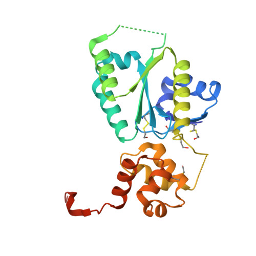
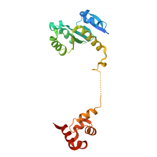
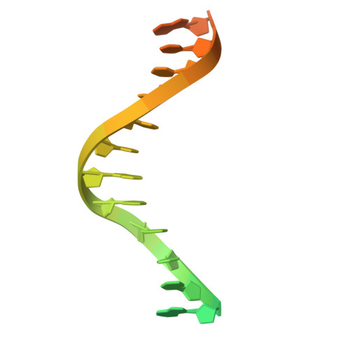
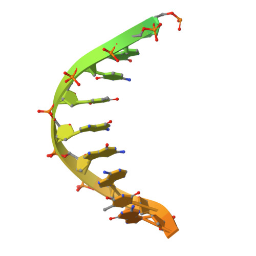
    FANCM is crucial in genome maintenance, functioning in the Fanconi anemia (FA) pathway, alternative lengthening of telomeres (ALT), and replication fork protection. FANCM recognizes branched DNA structures and promotes their remodeling through ATP-dependent branch migration. The protein has emerged as a promising therapeutic target due to synthetic lethal interactions with BRCA1, SMARCAL1, and RAD52, and in ALT-positive cancers. Here we present crystal structures of FANCM's N-terminal ATP-dependent translocase domain (2.2 Å) and C-terminal FAAP24-bound region (2.4 Å), both complexed with branched DNA. Through structural analysis, biochemical reconstitution, and cellular studies, we demonstrate that FANCM employs two distinct mechanisms: an ATP-dependent branch migration activity essential for DNA damage survival, and a branched DNA-binding mode that enhances FANCD2-FANCI monoubiquitination through FA core complex interaction. The N-terminal translocase domain specifically recognizes DNA junctions through multiple key elements, while the C-terminal FAAP24-binding domain engages adjacent double-stranded DNA. Our results reveal how FANCM evolved from an ancient DNA repair motor into a sophisticated sensor that couples DNA damage recognition to selective pathway activation, providing a structural framework for developing targeted therapeutics.


  • Organizational Affiliation
    • Genome Stability Unit, St. Vincent's Institute of Medical Research, Fitzroy, VIC, Australia.

Macromolecules

Find similar proteins by:  (by identity cutoff)  |  3D Structure
Entity ID: 1
MoleculeChains Sequence LengthOrganismDetailsImage
Fanconi anemia group M protein
A, C
234Homo sapiensMutation(s): 0 
Gene Names: FANCMKIAA1596
EC: 3.6.4.13
UniProt & NIH Common Fund Data Resources
Find proteins for Q8IYD8 (Homo sapiens)
Explore Q8IYD8 
Go to UniProtKB:  Q8IYD8
PHAROS:  Q8IYD8
GTEx:  ENSG00000187790 
Entity Groups  
Sequence Clusters30% Identity50% Identity70% Identity90% Identity95% Identity100% Identity
UniProt GroupQ8IYD8
Sequence Annotations
Expand
  • Reference Sequence
Find similar proteins by:  (by identity cutoff)  |  3D Structure
Entity ID: 2
MoleculeChains Sequence LengthOrganismDetailsImage
Fanconi anemia core complex-associated protein 24
B, D
215Homo sapiensMutation(s): 0 
Gene Names: FAAP24C19orf40
UniProt & NIH Common Fund Data Resources
Find proteins for Q9BTP7 (Homo sapiens)
Explore Q9BTP7 
Go to UniProtKB:  Q9BTP7
PHAROS:  Q9BTP7
GTEx:  ENSG00000131944 
Entity Groups  
Sequence Clusters30% Identity50% Identity70% Identity90% Identity95% Identity100% Identity
UniProt GroupQ9BTP7
Sequence Annotations
Expand
  • Reference Sequence

Find similar nucleic acids by:  Sequence   |   3D Structure  

Entity ID: 3
MoleculeChains LengthOrganismImage
DNA (25-MER)E [auth F],
G [auth H]
25synthetic construct
Sequence Annotations
Expand
  • Reference Sequence

Find similar nucleic acids by:  Sequence   |   3D Structure  

Entity ID: 4
MoleculeChains LengthOrganismImage
DNA (25-MER)F [auth G],
H [auth I]
25synthetic construct
Sequence Annotations
Expand
  • Reference Sequence
Experimental Data & Validation

Experimental Data

  • Method: X-RAY DIFFRACTION
  • Resolution: 2.40 Å
  • R-Value Free:  0.265 (Depositor), 0.265 (DCC) 
  • R-Value Work:  0.214 (Depositor), 0.215 (DCC) 
Space Group: P 1
Unit Cell:
Length ( Å )Angle ( ˚ )
a = 61.008α = 91.629
b = 69.975β = 89.929
c = 86.76γ = 115.952
Software Package:
Software NamePurpose
REFMACrefinement
DIALSdata reduction
DIALSdata scaling
PHASERphasing

Structure Validation

View Full Validation Report


Entry History & Funding Information

Deposition Data


Funding OrganizationLocationGrant Number
National Health and Medical Research Council (NHMRC, Australia)AustraliaGNT1181110
The Francis Crick InstituteUnited KingdomCC2068

Revision History  (Full details and data files)

  • Version 1.0: 2025-06-11
    Type: Initial release
  • Version 1.1: 2025-07-23
    Changes: Database references