9HJ1 | pdb_00009hj1

Cryo-EM structure of CDK2-cyclin A bound to a SCAPER peptide


Experimental Data Snapshot

  • Method: ELECTRON MICROSCOPY
  • Resolution: 2.90 Å
  • Aggregation State: PARTICLE 
  • Reconstruction Method: SINGLE PARTICLE 

Starting Model: in silico
View more details

wwPDB Validation   3D Report Full Report


This is version 1.1 of the entry. See complete history


Literature

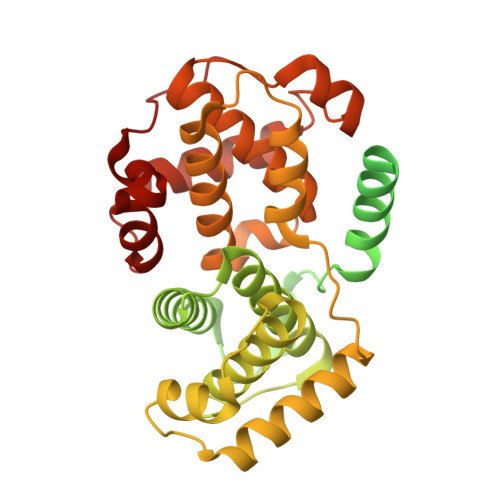
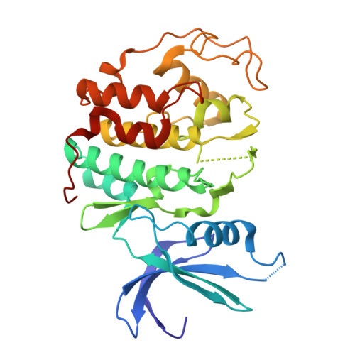
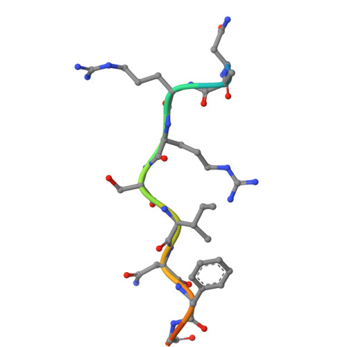
High-throughput investigation of cyclin docking interactions reveals the complexity of motif binding determinants.

Ord, M.Winters, M.J.Subbanna, M.S.de Martin Garrido, N.Cushing, V.I.Kliche, J.Benz, C.Ivarsson, Y.Greber, B.J.Pryciak, P.M.Davey, N.E.

(2025) Nat Commun 16: 7622-7622

  • DOI: https://doi.org/10.1038/s41467-025-62765-z
  • Primary Citation of Related Structures:  
    9HIU, 9HIW, 9HJ1

  • PubMed Abstract: 

    Many regulatory protein-protein interactions depend on Short Linear Motifs (SLiMs). In the cell cycle, cyclin-CDKs recognize SLiMs to control substrate recruitment and phosphorylation timing. Here, we measure the relative binding strength of ~100,000 peptides to 11 human cyclins from five families (D, E, A, B, and F). Using a quantitative intracellular binding assay and large-scale tiled peptide screening, we identify multiple non-canonical binders unveiling a broader repertoire of cyclin docking motif types. Cryo-electron microscopy and saturation mutagenesis studies reveal distinct binding modes and sequence features governing motif recognition, binding strength, and cyclin preference. Docking motifs vary from highly selective to pan-cyclin, thereby fine-tuning the timing of CDK phosphorylation during cell cycle. Overall, these findings provide insights into the rules encoding specificity and affinity of SLiM-mediated interactions and offer a framework for understanding motif-driven protein networks across the proteome.


  • Organizational Affiliation
    • University of Cambridge, CRUK Cambridge Institute, Cambridge, UK.

Macromolecules
Find similar proteins by:  (by identity cutoff)  |  3D Structure
Entity ID: 1
MoleculeChains Sequence LengthOrganismDetailsImage
Cyclin-A2432Homo sapiensMutation(s): 1 
Gene Names: CCNA2CCN1CCNA
UniProt & NIH Common Fund Data Resources
Find proteins for P20248 (Homo sapiens)
Explore P20248 
Go to UniProtKB:  P20248
PHAROS:  P20248
GTEx:  ENSG00000145386 
Entity Groups  
Sequence Clusters30% Identity50% Identity70% Identity90% Identity95% Identity100% Identity
UniProt GroupP20248
Sequence Annotations
Expand
  • Reference Sequence
Find similar proteins by:  (by identity cutoff)  |  3D Structure
Entity ID: 2
MoleculeChains Sequence LengthOrganismDetailsImage
Cyclin-dependent kinase 2297Homo sapiensMutation(s): 0 
Gene Names: CDK2CDKN2
EC: 2.7.11.22
UniProt & NIH Common Fund Data Resources
Find proteins for P24941 (Homo sapiens)
Explore P24941 
Go to UniProtKB:  P24941
PHAROS:  P24941
GTEx:  ENSG00000123374 
Entity Groups  
Sequence Clusters30% Identity50% Identity70% Identity90% Identity95% Identity100% Identity
UniProt GroupP24941
Sequence Annotations
Expand
  • Reference Sequence

Find similar proteins by:  Sequence   |   3D Structure  

Entity ID: 3
MoleculeChains Sequence LengthOrganismDetailsImage
SCAPER13Homo sapiensMutation(s): 0 
UniProt & NIH Common Fund Data Resources
Find proteins for Q9BY12 (Homo sapiens)
Explore Q9BY12 
Go to UniProtKB:  Q9BY12
PHAROS:  Q9BY12
GTEx:  ENSG00000140386 
Entity Groups  
Sequence Clusters30% Identity50% Identity70% Identity90% Identity95% Identity100% Identity
UniProt GroupQ9BY12
Sequence Annotations
Expand
  • Reference Sequence
Experimental Data & Validation

Experimental Data

  • Method: ELECTRON MICROSCOPY
  • Resolution: 2.90 Å
  • Aggregation State: PARTICLE 
  • Reconstruction Method: SINGLE PARTICLE 
EM Software:
TaskSoftware PackageVersion
MODEL REFINEMENTPHENIX1.21.1_5286
RECONSTRUCTIONRELION5.0 beta

Structure Validation

View Full Validation Report


Entry History & Funding Information

Deposition Data


Funding OrganizationLocationGrant Number
Not funded--

Revision History  (Full details and data files)

  • Version 1.0: 2025-08-20
    Type: Initial release
  • Version 1.1: 2026-03-04
    Changes: Data collection, Database references