Compounds mimicking the Michaelis-Menten transition state of the phosphatidylinositol 4-kinase.
Hrebabecky, H., Klima, M., Dejmek, M., Krupa, P., Fernandez, M.R., Davidova, E., Benysek, J., Martinez-Seara, H., Rozycki, B., Boura, E., Nencka, R.(2026) Bioorg Med Chem 139: 118666-118666
- PubMed: 42086018 Search on PubMed
- DOI: https://doi.org/10.1016/j.bmc.2026.118666
- Primary Citation Related Structures:
9HHM - PubMed Abstract:
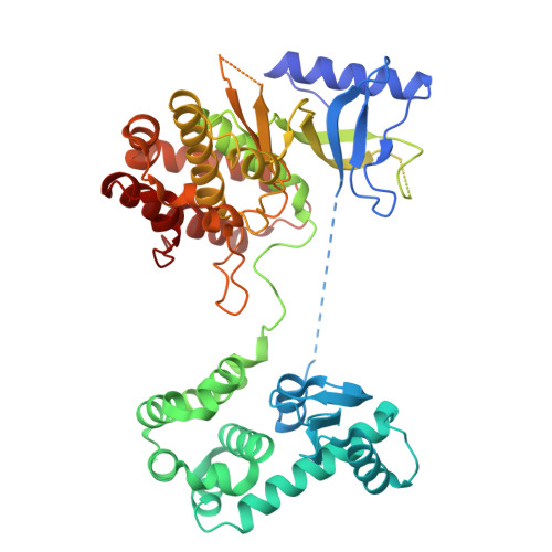
Phosphatidylinositol 4-kinases (PI4Ks) are crucial enzymes in lipid signaling responsible for generating phosphatidylinositol-4-phosphate (PI4P). Although the ATP-binding site of PI4Ks has been extensively studied, the structural characterization of their natural substrate, phosphatidylinositol (PI), bound to the enzyme remains elusive. In this study, we synthesized novel non-hydrolyzable Michaelis-Menten transition state mimetics in which the ADP molecule is covalently linked to inositol-4-phosphate or simple phosphatidylinositol-4-phosphate via a methylene or ethylene bridge. During our synthesis efforts, we successfully addressed the significantly limited reactivity at position 4 of inositols by utilizing P(III) phosphorus reagents, which proved crucial for the synthesis of these mimetics. For the simplest ADP-C-4PI analogue, we obtained a crystal structure of PI4K2B, which can be used to understand how these phospholipids are phosphorylated on membrane surfaces.
- Institute of Organic Chemistry and Biochemistry, Academy of Sciences of the Czech Republic, v.v.i, Flemingovo náměstí 542/2, 166 10 Prague, Czech Republic.
Organizational Affiliation: