9HAN | pdb_00009han

Bovine collagen VI local refinement of C-terminal region


Experimental Data Snapshot

  • Method: ELECTRON MICROSCOPY
  • Resolution: 4.33 Å
  • Aggregation State: PARTICLE 
  • Reconstruction Method: SINGLE PARTICLE 

wwPDB Validation   3D Report Full Report


This is version 1.1 of the entry. See complete history


Literature

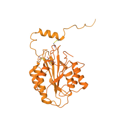
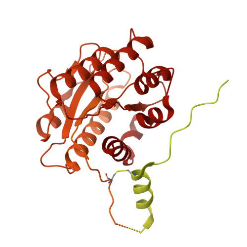
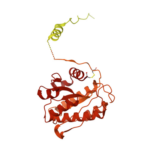
Collagen VI microfibril structure reveals mechanism for molecular assembly and clustering of inherited pathogenic mutations.

Godwin, A.R.F.Becker, M.H.Dajani, R.Snee, M.Roseman, A.M.Baldock, C.

(2025) Nat Commun 16: 7549-7549

  • DOI: https://doi.org/10.1038/s41467-025-62923-3
  • Primary Citation of Related Structures:  
    9GTU, 9HAN

  • PubMed Abstract: 

    Collagen VI links the cell surface to the extracellular matrix to provide mechanical strength to most mammalian tissues, and is linked to human diseases including muscular dystrophy, fibrosis, cardiovascular disease and osteoarthritis. Collagen VI assembles from heterotrimers of three different α-chains into microfibrils, but there are many gaps in our knowledge of the molecular assembly process. Here, we determine the structures of both heterotrimeric mini-collagen VI constructs and collagen VI microfibrils, from mammalian tissue, using cryogenic-electron microscopy. These structures reveal a cysteine-rich coiled coil region involved in trimerisation as well as microfibril assembly. Furthermore, our structures show that pathogenic mutations are located at interaction sites involved in different steps of collagen VI assembly, from the trimeric-coiled coil region that mediates heterotrimerisation, to clusters of mutations in the triple-helical region involved in microfibril formation. Our microfibril structure provides a template for understanding supramolecular assembly, and offers a platform for rationale design of therapeutics for collagen VI pathologies.


  • Organizational Affiliation
    • Division of Cell-Matrix Biology and Regenerative Medicine, Manchester Cell-Matrix Centre, School of Biological Sciences, Faculty of Biology, Medicine and Health, Manchester Academic Health Science Centre, University of Manchester, Manchester, UK.

Macromolecules
Find similar proteins by:  (by identity cutoff)  |  3D Structure
Entity ID: 1
MoleculeChains Sequence LengthOrganismDetailsImage
Collagen type VI alpha 3 chainA [auth C],
D [auth F]
3,061Bos taurusMutation(s): 0 
UniProt
Find proteins for A0AAA9TAB4 (Bos taurus)
Explore A0AAA9TAB4 
Go to UniProtKB:  A0AAA9TAB4
Entity Groups  
Sequence Clusters30% Identity50% Identity70% Identity90% Identity95% Identity100% Identity
UniProt GroupA0AAA9TAB4
Glycosylation
Glycosylation Sites: 1
Sequence Annotations
Expand
  • Reference Sequence
Find similar proteins by:  (by identity cutoff)  |  3D Structure
Entity ID: 2
MoleculeChains Sequence LengthOrganismDetailsImage
Collagen type VI alpha 1 chainB [auth A],
E [auth D]
1,027Bos taurusMutation(s): 0 
UniProt
Find proteins for E1BI98 (Bos taurus)
Explore E1BI98 
Go to UniProtKB:  E1BI98
Entity Groups  
Sequence Clusters30% Identity50% Identity70% Identity90% Identity95% Identity100% Identity
UniProt GroupE1BI98
Glycosylation
Glycosylation Sites: 1
Sequence Annotations
Expand
  • Reference Sequence
Find similar proteins by:  (by identity cutoff)  |  3D Structure
Entity ID: 3
MoleculeChains Sequence LengthOrganismDetailsImage
Collagen type VI alpha 2 chainC [auth B],
F [auth E]
1,027Bos taurusMutation(s): 0 
UniProt
Find proteins for A0AAA9TXG2 (Bos taurus)
Explore A0AAA9TXG2 
Go to UniProtKB:  A0AAA9TXG2
Entity Groups  
Sequence Clusters30% Identity50% Identity70% Identity90% Identity95% Identity100% Identity
UniProt GroupA0AAA9TXG2
Glycosylation
Glycosylation Sites: 2
Sequence Annotations
Expand
  • Reference Sequence
Oligosaccharides

Help

Entity ID: 4
MoleculeChains Length2D Diagram Glycosylation3D Interactions
2-acetamido-2-deoxy-beta-D-glucopyranose-(1-4)-[alpha-L-fucopyranose-(1-6)]2-acetamido-2-deoxy-beta-D-glucopyranose
G, J
3N-Glycosylation
Glycosylation Resources
GlyTouCan:  G21290RB
GlyCosmos:  G21290RB
GlyGen:  G21290RB
Entity ID: 5
MoleculeChains Length2D Diagram Glycosylation3D Interactions
2-acetamido-2-deoxy-beta-D-glucopyranose-(1-4)-2-acetamido-2-deoxy-beta-D-glucopyranose
H, I, K
2N-Glycosylation
Glycosylation Resources
GlyTouCan:  G42666HT
GlyCosmos:  G42666HT
GlyGen:  G42666HT
Entity ID: 6
MoleculeChains Length2D Diagram Glycosylation3D Interactions
beta-D-mannopyranose-(1-4)-2-acetamido-2-deoxy-beta-D-glucopyranose-(1-4)-2-acetamido-2-deoxy-beta-D-glucopyranose
L
3N-Glycosylation
Glycosylation Resources
GlyTouCan:  G15407YE
GlyCosmos:  G15407YE
GlyGen:  G15407YE
Experimental Data & Validation

Experimental Data

  • Method: ELECTRON MICROSCOPY
  • Resolution: 4.33 Å
  • Aggregation State: PARTICLE 
  • Reconstruction Method: SINGLE PARTICLE 
EM Software:
TaskSoftware PackageVersion
MODEL REFINEMENTPHENIX

Structure Validation

View Full Validation Report


Entry History & Funding Information

Deposition Data


Funding OrganizationLocationGrant Number
Biotechnology and Biological Sciences Research Council (BBSRC)United Kingdom--

Revision History  (Full details and data files)

  • Version 1.0: 2025-08-20
    Type: Initial release
  • Version 1.1: 2025-08-27
    Changes: Data collection, Database references