9H9R | pdb_00009h9r

Full gamma-tubulin ring complex composed of the Candida albicans gamma-tubulin small complex in complex with Spc72 CM1


Experimental Data Snapshot

  • Method: ELECTRON MICROSCOPY
  • Resolution: 8.20 Å
  • Aggregation State: PARTICLE 
  • Reconstruction Method: SINGLE PARTICLE 

wwPDB Validation   3D Report Full Report


This is version 1.0 of the entry. See complete history


Literature

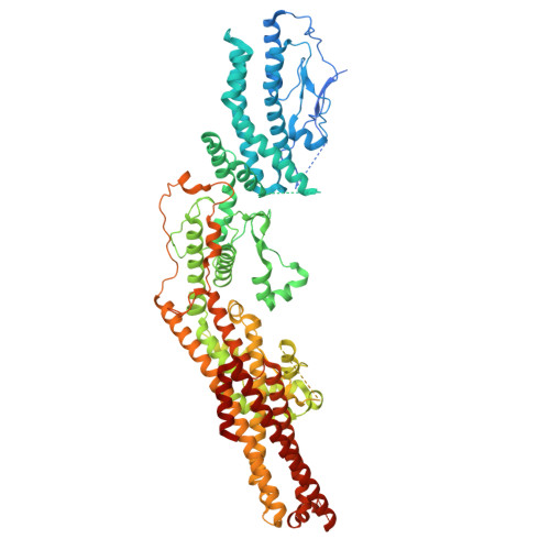
Structural insights into the interplay between microtubule polymerases, gamma-tubulin complexes and their receptors.

Zheng, A.Vermeulen, B.J.A.Wurtz, M.Neuner, A.Lubbehusen, N.Mayer, M.P.Schiebel, E.Pfeffer, S.

(2025) Nat Commun 16: 402-402

  • DOI: https://doi.org/10.1038/s41467-024-55778-7
  • Primary Citation of Related Structures:  
    9H9P, 9H9Q, 9H9R

  • PubMed Abstract: 

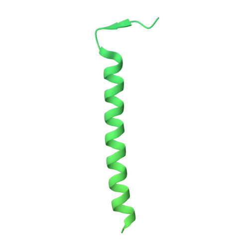
    The γ-tubulin ring complex (γ-TuRC) is a structural template for controlled nucleation of microtubules from α/β-tubulin heterodimers. At the cytoplasmic side of the yeast spindle pole body, the CM1-containing receptor protein Spc72 promotes γ-TuRC assembly from seven γ-tubulin small complexes (γ-TuSCs) and recruits the microtubule polymerase Stu2, yet their molecular interplay remains unclear. Here, we determine the cryo-EM structure of the Candida albicans cytoplasmic nucleation unit at 3.6 Å resolution, revealing how the γ-TuRC is assembled and conformationally primed for microtubule nucleation by the dimerised Spc72 CM1 motif. Two coiled-coil regions of Spc72 interact with the conserved C-terminal α-helix of Stu2 and thereby position the α/β-tubulin-binding TOG domains of Stu2 in the vicinity of the microtubule assembly site. Collectively, we reveal the function of CM1 motifs in γ-TuSC oligomerisation and the recruitment of microtubule polymerases to the γ-TuRC.


  • Organizational Affiliation
    • Zentrum für Molekulare Biologie der Universität Heidelberg (ZMBH), Heidelberg, Germany.

Macromolecules
Find similar proteins by:  (by identity cutoff)  |  3D Structure
Entity ID: 1
MoleculeChains Sequence LengthOrganismDetailsImage
Tubulin gamma chain498Candida albicansMutation(s): 0 
Gene Names: FOB64_001277
UniProt
Find proteins for A0A1D8PMD4 (Candida albicans (strain SC5314 / ATCC MYA-2876))
Explore A0A1D8PMD4 
Go to UniProtKB:  A0A1D8PMD4
Entity Groups  
Sequence Clusters30% Identity50% Identity70% Identity90% Identity95% Identity100% Identity
UniProt GroupA0A1D8PMD4
Sequence Annotations
Expand
  • Reference Sequence
Find similar proteins by:  (by identity cutoff)  |  3D Structure
Entity ID: 2
MoleculeChains Sequence LengthOrganismDetailsImage
Spindle pole body component896Candida albicansMutation(s): 0 
Gene Names: CAALFM_CR06740WAorf19.8327
UniProt
Find proteins for Q59PZ2 (Candida albicans (strain SC5314 / ATCC MYA-2876))
Explore Q59PZ2 
Go to UniProtKB:  Q59PZ2
Entity Groups  
Sequence Clusters30% Identity50% Identity70% Identity90% Identity95% Identity100% Identity
UniProt GroupQ59PZ2
Sequence Annotations
Expand
  • Reference Sequence
Find similar proteins by:  (by identity cutoff)  |  3D Structure
Entity ID: 3
MoleculeChains Sequence LengthOrganismDetailsImage
Spc98p810Candida albicansMutation(s): 0 
Gene Names: SPC98CAALFM_CR01990CAorf19.10131
UniProt
Find proteins for A0A1D8PS42 (Candida albicans (strain SC5314 / ATCC MYA-2876))
Explore A0A1D8PS42 
Go to UniProtKB:  A0A1D8PS42
Entity Groups  
Sequence Clusters30% Identity50% Identity70% Identity90% Identity95% Identity100% Identity
UniProt GroupA0A1D8PS42
Sequence Annotations
Expand
  • Reference Sequence
Find similar proteins by:  (by identity cutoff)  |  3D Structure
Entity ID: 4
MoleculeChains Sequence LengthOrganismDetailsImage
Mto2p-binding domain-containing protein615Candida albicansMutation(s): 0 
Gene Names: CAALFM_C701460CAorf19.13936
UniProt
Find proteins for Q5AGV5 (Candida albicans (strain SC5314 / ATCC MYA-2876))
Explore Q5AGV5 
Go to UniProtKB:  Q5AGV5
Entity Groups  
Sequence Clusters30% Identity50% Identity70% Identity90% Identity95% Identity100% Identity
UniProt GroupQ5AGV5
Sequence Annotations
Expand
  • Reference Sequence
Experimental Data & Validation

Experimental Data

  • Method: ELECTRON MICROSCOPY
  • Resolution: 8.20 Å
  • Aggregation State: PARTICLE 
  • Reconstruction Method: SINGLE PARTICLE 
EM Software:
TaskSoftware PackageVersion
RECONSTRUCTIONRELION3.0

Structure Validation

View Full Validation Report


Entry History & Funding Information

Deposition Data


Funding OrganizationLocationGrant Number
German Research Foundation (DFG)GermanySchi 295/4-4
German Research Foundation (DFG)GermanyPF 963/1-4
German Research Foundation (DFG)GermanyPF 963/4-1
Other private--
German Research Foundation (DFG)GermanyINST 35/1314-1 FUGG
German Research Foundation (DFG)GermanyINST 35/1134-1 FUGG

Revision History  (Full details and data files)

  • Version 1.0: 2025-01-15
    Type: Initial release