9H25 | pdb_00009h25

Structure of rsCherry exposed to oxygen for 16 days


Experimental Data Snapshot

  • Method: X-RAY DIFFRACTION
  • Resolution: 1.90 Å
  • R-Value Free: 
    0.190 (Depositor), 0.191 (DCC) 
  • R-Value Work: 
    0.156 (Depositor), 0.156 (DCC) 
  • R-Value Observed: 
    0.158 (Depositor) 

Starting Model: experimental
View more details

wwPDB Validation   3D Report Full Report


This is version 1.0 of the entry. See complete history


Literature

Crystal structure analysis of oxygen-induced degradation occurring in rsCherry.

Bui, T.Y.H.Pecqueur, L.Dedecker, P.Van Meervelt, L.

(2025) Acta Crystallogr F Struct Biol Commun 81: 311-318

  • DOI: https://doi.org/10.1107/S2053230X25005485
  • Primary Citation of Related Structures:  
    9H25, 9H26, 9H27, 9JX2

  • PubMed Abstract: 

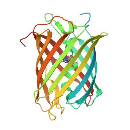
    rsCherry was one of the first reversibly photoswitchable variants to be developed from mCherry. However, its practical applications have been limited due to several inherent drawbacks. We have recently shown that the purified protein undergoes oxygen-induced chromophore degradation in solution, resulting in the progressive loss of its fluorescence and color. In this work, we present four crystal structures of rsCherry that exhibit varying degrees of degradation. Our structural analysis indicates that oxygen-induced degradation of rsCherry predominantly affects the chromophore without altering the protein backbone. Changes were only observed in the conformation of Lys70, confirming the crucial role of this residue in chromophore damage in rsCherry. Overall, this study provides valuable insights into the structural changes triggered by oxygen exposure in rsCherry, offering suggestions for the development of stable red fluorescent proteins with improved resistance to oxidative damage.


  • Organizational Affiliation
    • Biochemistry, Molecular and Structural Biology, Department of Chemistry, KU Leuven, Leuven, Belgium.

Macromolecules
Find similar proteins by:  (by identity cutoff)  |  3D Structure
Entity ID: 1
MoleculeChains Sequence LengthOrganismDetailsImage
rsCherry263Discosoma sp.Mutation(s): 32 
Gene Names: PAmCherryPAmCherry1
UniProt
Find proteins for Q9U6Y8 (Discosoma sp.)
Explore Q9U6Y8 
Go to UniProtKB:  Q9U6Y8
Entity Groups  
Sequence Clusters30% Identity50% Identity70% Identity90% Identity95% Identity100% Identity
UniProt GroupQ9U6Y8
Sequence Annotations
Expand
  • Reference Sequence
Small Molecules
Modified Residues  3 Unique
IDChains TypeFormula2D DiagramParent
Q2K
Query on Q2K
A
L-PEPTIDE LINKINGC9 H11 N3 O4 SMET, TYR, GLY
QIP
Query on QIP
A
L-PEPTIDE LINKINGC9 H14 N4 O4 SMET, TYR, GLY
QYX
Query on QYX
A
L-PEPTIDE LINKINGC16 H18 N3 O4 SMET, TYR, GLY
Experimental Data & Validation

Experimental Data

  • Method: X-RAY DIFFRACTION
  • Resolution: 1.90 Å
  • R-Value Free:  0.190 (Depositor), 0.191 (DCC) 
  • R-Value Work:  0.156 (Depositor), 0.156 (DCC) 
  • R-Value Observed: 0.158 (Depositor) 
Space Group: I 1 2 1
Unit Cell:
Length ( Å )Angle ( ˚ )
a = 61.391α = 90
b = 43.452β = 106
c = 108.818γ = 90
Software Package:
Software NamePurpose
PHENIXrefinement
Aimlessdata scaling
DIALSdata reduction
PHASERphasing

Structure Validation

View Full Validation Report


Entry History & Funding Information

Deposition Data


Funding OrganizationLocationGrant Number
KU LeuvenBelgium--

Revision History  (Full details and data files)

  • Version 1.0: 2025-08-20
    Type: Initial release