9GYH | pdb_00009gyh

HEW Lysozyme with His 15 functionalized with iodoacetamide


Experimental Data Snapshot

  • Method: X-RAY DIFFRACTION
  • Resolution: 1.60 Å
  • R-Value Free: 
    0.211 (Depositor), 0.210 (DCC) 
  • R-Value Work: 
    0.173 (Depositor), 0.173 (DCC) 
  • R-Value Observed: 
    0.175 (Depositor) 

Starting Model: experimental
View more details

wwPDB Validation 3D Report Full Report

Validation slider image for 9GYH

This is version 1.0 of the entry. See complete history

Literature

The structure of His15 acetamide-modified hen egg-white lysozyme: a nice surprise from an old friend.

Malanho da Silva, J.Lanuza, J.Bruno, F.Calderone, V.Ravera, E.

(2025) Acta Crystallogr F Struct Biol Commun 81: 41-46

  • DOI: https://doi.org/10.1107/S2053230X2500010X
  • Primary Citation Related Structures: 
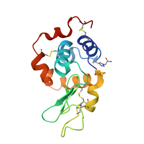
    9GYH

  • PubMed Abstract: 

    Hen egg-white lysozyme (HEWL) is a small polycationic protein which is highly soluble and stable. This has led to it becoming a `molecular laboratory' where chemical biological operations and structural techniques are tested. To date, HEWL accounts for 1233 PDB entries, roughly 0.5% of the total, making it the best-represented protein in the PDB. With the aim of unambiguously identifying the N atom of the His15 side chain that is most reactive towards iodoacetamide, the structure of chemically modified HEWL was determined by crystallizing it using the `15 minutes lysozyme' protocol. This protocol invariably yields tetragonal crystals of the unmodified protein. To our surprise, we found that the crystals of the modified protein had similar unit-cell parameters but that refinement was only possible when considering an orthorhombic system.


  • Organizational Affiliation
    • Department of Chemistry `Ugo Schiff', Università degli Studi di Firenze, Via della Lastruccia 3, 50019 Sesto Fiorentino, Italy.

Macromolecule Content 

  • Total Structure Weight: 29.1 kDa 
  • Atom Count: 2,420 
  • Modeled Residue Count: 258 
  • Deposited Residue Count: 258 
  • Unique protein chains: 1

Macromolecules

Find similar proteins by:|  3D Structure
Entity ID: 1
MoleculeChains  Sequence LengthOrganismDetailsImage
Lysozyme C
A, B
129Gallus gallusMutation(s): 0 
Gene Names: LYZ
EC: 3.2.1.17
UniProt
Find proteins for P00698 (Gallus gallus)
Explore P00698 
Go to UniProtKB:  P00698
Entity Groups
Sequence Clusters30% Identity50% Identity70% Identity90% Identity95% Identity100% Identity
UniProt GroupP00698
Sequence Annotations
Expand
Reference Sequence

Small Molecules

Ligands 2 Unique
IDChains Name / Formula / InChI Key2D Diagram3D Interactions
CL

Query on CL


Download:Ideal Coordinates CCD File
D [auth A]
E [auth A]
F [auth A]
G [auth A]
I [auth B]
D [auth A],
E [auth A],
F [auth A],
G [auth A],
I [auth B],
J [auth B],
K [auth B],
L [auth B]
CHLORIDE ION
Cl
VEXZGXHMUGYJMC-UHFFFAOYSA-M
NA

Query on NA


Download:Ideal Coordinates CCD File
C [auth A],
H [auth B]
SODIUM ION
Na
FKNQFGJONOIPTF-UHFFFAOYSA-N
Modified Residues  1 Unique
IDChains TypeFormula2D DiagramParent
A1IQX
Query on A1IQX
A, B
L-PEPTIDE LINKINGC8 H12 N4 O3HIS

Experimental Data & Validation

Experimental Data

  • Method: X-RAY DIFFRACTION
  • Resolution: 1.60 Å
  • R-Value Free:  0.211 (Depositor), 0.210 (DCC) 
  • R-Value Work:  0.173 (Depositor), 0.173 (DCC) 
  • R-Value Observed: 0.175 (Depositor) 
Space Group: P 21 21 21
Unit Cell:
Length ( Å )Angle ( ˚ )
a = 37.53α = 90
b = 78.17β = 90
c = 79.31γ = 90
Software Package:
Software NamePurpose
PHENIXrefinement
XDSdata reduction
XSCALEdata scaling
MOLREPphasing

Structure Validation

View Full Validation Report


Entry History 

& Funding Information

Deposition Data


Funding OrganizationLocationGrant Number
Italian Ministry of EducationItaly--

Revision History  (Full details and data files)

  • Version 1.0: 2025-02-26
    Type: Initial release