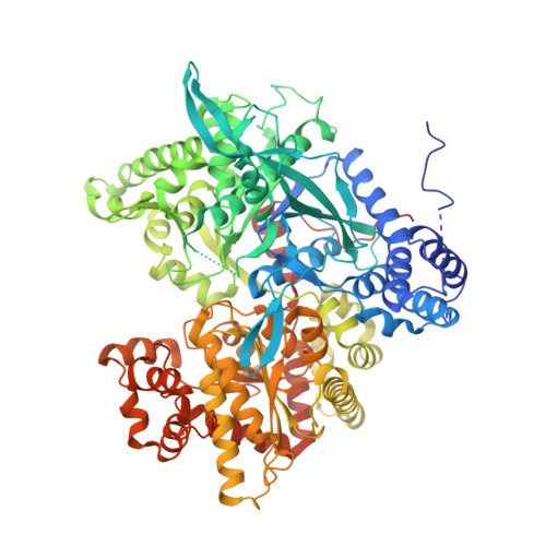
9GPB

THE ALLOSTERIC TRANSITION OF GLYCOGEN PHOSPHORYLASE


Experimental Data Snapshot

  • Method: X-RAY DIFFRACTION
  • Resolution: 2.90 Å
  • R-Value Work: 0.177 

wwPDB Validation   3D Report Full Report


This is version 1.3 of the entry. See complete history


Literature

The allosteric transition of glycogen phosphorylase.

Barford, D.Johnson, L.N.

(1989) Nature 340: 609-616

  • DOI: https://doi.org/10.1038/340609a0
  • Primary Citation of Related Structures:  
    9GPB

  • PubMed Abstract: 

    The crystal structure of R-state glycogen phosphorylase b has been determined at 2.9 A resolution. A comparison of T-state and R-state structures of the enzyme explains its cooperative behaviour on ligand binding and the allosteric regulation of its activity. Communication between catalytic sites of the dimer is provided by a change in packing geometry of two helices linking each site with the subunit interface. Activation by AMP or by phosphorylation results in a quaternary conformational change that switches these two helices into the R-state conformation.


  • Organizational Affiliation

    Laboratory of Molecular Biophysics, University of Oxford, UK.


Macromolecules
Find similar proteins by:  (by identity cutoff)  |  3D Structure
Entity ID: 1
MoleculeChains Sequence LengthOrganismDetailsImage
GLYCOGEN PHOSPHORYLASE B
A, B, C, D
842Oryctolagus cuniculusMutation(s): 0 
EC: 2.4.1.1
UniProt
Find proteins for P00489 (Oryctolagus cuniculus)
Explore P00489 
Go to UniProtKB:  P00489
Entity Groups  
Sequence Clusters30% Identity50% Identity70% Identity90% Identity95% Identity100% Identity
UniProt GroupP00489
Sequence Annotations
Expand
  • Reference Sequence
Small Molecules
Ligands 2 Unique
IDChains Name / Formula / InChI Key2D Diagram3D Interactions
PLP
Query on PLP

Download Ideal Coordinates CCD File 
H [auth A],
L [auth B],
P [auth C],
T [auth D]
PYRIDOXAL-5'-PHOSPHATE
C8 H10 N O6 P
NGVDGCNFYWLIFO-UHFFFAOYSA-N
SO4
Query on SO4

Download Ideal Coordinates CCD File 
E [auth A]
F [auth A]
G [auth A]
I [auth B]
J [auth B]
E [auth A],
F [auth A],
G [auth A],
I [auth B],
J [auth B],
K [auth B],
M [auth C],
N [auth C],
O [auth C],
Q [auth D],
R [auth D],
S [auth D]
SULFATE ION
O4 S
QAOWNCQODCNURD-UHFFFAOYSA-L
Experimental Data & Validation

Experimental Data

  • Method: X-RAY DIFFRACTION
  • Resolution: 2.90 Å
  • R-Value Work: 0.177 
  • Space Group: P 1 21 1
Unit Cell:
Length ( Å )Angle ( ˚ )
a = 119α = 90
b = 190β = 109.35
c = 88.2γ = 90
Software Package:
Software NamePurpose
X-PLORrefinement

Structure Validation

View Full Validation Report


Entry History 

Deposition Data

Revision History  (Full details and data files)

  • Version 1.0: 1992-10-15
    Type: Initial release
  • Version 1.1: 2008-03-25
    Changes: Version format compliance
  • Version 1.2: 2011-07-13
    Changes: Non-polymer description, Version format compliance
  • Version 1.3: 2017-11-29
    Changes: Derived calculations, Other