Protocol for high-yield bacterial expression and purification of the voltage-dependent anion channel 1 for high-throughput biophysical assays.
Conti Nibali, S., Magri, A., Messina, A., Wagner, A., Duman, R., De Pinto, V., Turato, C., Arrigoni, C., Lolicato, M.(2025) STAR Protoc 6: 103557-103557
- PubMed: 39799574 Search on PubMed
- DOI: https://doi.org/10.1016/j.xpro.2024.103557
- Primary Citation Related Structures:
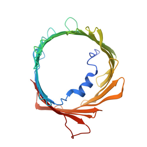
9GNG - PubMed Abstract:
Voltage-dependent anion channel 1 (VDAC1) is a key protein in cellular metabolism and apoptosis. Here, we present a protocol to express and purify milligram amounts of recombinant VDAC1 in Escherichia coli. We detail steps for a fluorescence polarization-based high-throughput screening assay using NADH displacement, along with procedures for thermostability, fluorescence polarization, and X-ray crystallography. In this context, we demonstrate how 2-methyl-2,4-pentanediol (MPD), a crystallization reagent, interferes with VDAC1 small-molecule binding, hindering the detection of these ligands in the crystal. For complete details on the use and execution of this protocol, please refer to Conti Nibali et al. 1 .
- Department of Molecular Medicine, University of Pavia, Pavia, Italy.
Organizational Affiliation: