


Crystal structure of human cathepsin S produced in insect cells in complex with ketoamide 13b
Falke, S., Karnicar, K., Usenik, A., Lindic, N., Sekirnik, A., Reinke, P.Y.A., Guenther, S., Turk, D., Meents, A.To be published.
Experimental Data Snapshot
Starting Model: experimental
View more details
Entity ID: 1 | |||||
|---|---|---|---|---|---|
| Molecule | Chains | Sequence Length | Organism | Details | Image |
| Cathepsin S | 242 | Homo sapiens | Mutation(s): 0 Gene Names: CTSS EC: 3.4.22.27 | 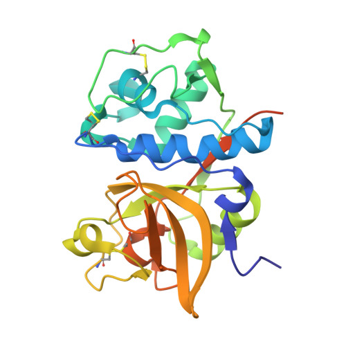 | |
UniProt & NIH Common Fund Data Resources | |||||
PHAROS: P25774 GTEx: ENSG00000163131 | |||||
Entity Groups | |||||
| Sequence Clusters | 30% Identity50% Identity70% Identity90% Identity95% Identity100% Identity | ||||
| UniProt Group | P25774 | ||||
Sequence AnnotationsExpand | |||||
Reference Sequence | |||||
| Ligands 4 Unique | |||||
|---|---|---|---|---|---|
| ID | Chains | Name / Formula / InChI Key | 2D Diagram | 3D Interactions | |
| KH0 (Subject of Investigation/LOI) Download:Ideal Coordinates CCD File | C [auth A], H [auth B] | ~{tert}-butyl ~{N}-[1-[(2~{S})-3-cyclopropyl-1-oxidanylidene-1-[[(2~{S},3~{S})-3-oxidanyl-4-oxidanylidene-1-[(3~{S})-2-oxidanylidenepyrrolidin-3-yl]-4-[(phenylmethyl)amino]butan-2-yl]amino]propan-2-yl]-2-oxidanylidene-pyridin-3-yl]carbamate C31 H41 N5 O7 FRACPXUHUTXLCX-LFBFJMOVSA-N |  | ||
| CIT Download:Ideal Coordinates CCD File | J [auth B] | CITRIC ACID C6 H8 O7 KRKNYBCHXYNGOX-UHFFFAOYSA-N |  | ||
| DMS Download:Ideal Coordinates CCD File | F [auth A], G [auth A] | DIMETHYL SULFOXIDE C2 H6 O S IAZDPXIOMUYVGZ-UHFFFAOYSA-N |  | ||
| EDO Download:Ideal Coordinates CCD File | D [auth A], E [auth A], I [auth B], K [auth B] | 1,2-ETHANEDIOL C2 H6 O2 LYCAIKOWRPUZTN-UHFFFAOYSA-N |  | ||
| Length ( Å ) | Angle ( ˚ ) |
|---|---|
| a = 86.102 | α = 90 |
| b = 86.102 | β = 90 |
| c = 152.045 | γ = 90 |
| Software Name | Purpose |
|---|---|
| PHENIX | refinement |
| XDS | data reduction |
| XSCALE | data scaling |
| PHENIX | phasing |
| Funding Organization | Location | Grant Number |
|---|---|---|
| Helmholtz Association | Germany | FISCOV |
| Helmholtz Association | Germany | SFragX |
| German Federal Ministry for Education and Research | Germany | 031B0405 |
| Slovenian Research Agency | Slovenia | P1-0048 |
| Slovenian Research Agency | Slovenia | IO-0048 |


