9GFK | pdb_00009gfk

human MDM2 complex with stapled foldamer


Experimental Data Snapshot

  • Method: X-RAY DIFFRACTION
  • Resolution: 1.84 Å
  • R-Value Free: 
    0.239 (Depositor), 0.234 (DCC) 
  • R-Value Work: 
    0.200 (Depositor), 0.193 (DCC) 
  • R-Value Observed: 
    0.202 (Depositor) 

Starting Model: experimental
View more details

wwPDB Validation   3D Report Full Report


Ligand Structure Quality Assessment 


This is version 1.2 of the entry. See complete history


Literature

Optimal Stapling of a Helical Peptide-Foldamer Hybrid Using a C-Terminal 4-Mercaptoproline Enhances Protein Surface Recognition and Cellular Activity.

Neuville, M.Bourgeais, M.Buratto, J.Saragaglia, C.Li, B.Galeano-Otero, I.Mauran, L.Varajao, L.Goudreau, S.R.Kauffmann, B.Thinon, E.Pasco, M.Khatib, A.M.Guichard, G.

(2025) Chemistry 31: e202403330-e202403330

  • DOI: https://doi.org/10.1002/chem.202403330
  • Primary Citation of Related Structures:  
    9GFK

  • PubMed Abstract: 

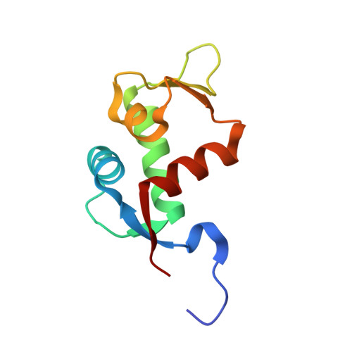
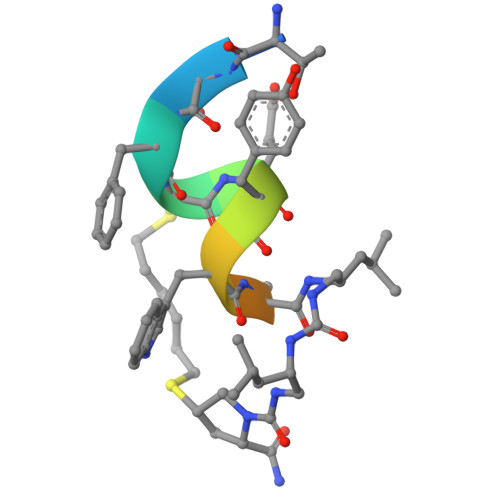
    Structural analysis of a co-crystal of a helically-folded peptide-foldamer hybrid in complex with hDM2 E3 ubiquitin ligase, revealed a unique orientation for the C-terminal proline with the pyrrolidine ring pointing backwards in the sequence, and suggested new opportunities for macrocyclization. In particular, we found that the C-terminal prolyl residue could be replaced by its (2S,4S)-4-mercaptoprolyl analogue for optimal bisthioether crosslinking with a cysteine residue installed at position 4 in the sequence. The resulting i,i+7 stapled peptide-foldamer is a high-affinity binder to hDM2, is cell permeable and restores the p53 signalling pathway in p53wt cancer cells. The co-crystal structure of hDM2 and the stapled peptide-foldamer hybrid was determined at 1.84 Å, fully validating the original design and further highlighting the potential of cis-4-mercaptoproline in the context of peptide and foldamer stapling.


  • Organizational Affiliation
    • Univ. Bordeaux, CNRS, Bordeaux INP, CBMN, UMR 5248, IECB, 2 rue Robert Escarpit, F-33600, Pessac, France.

Macromolecules
Find similar proteins by:  (by identity cutoff)  |  3D Structure
Entity ID: 1
MoleculeChains Sequence LengthOrganismDetailsImage
E3 ubiquitin-protein ligase Mdm2
A, B, C, D
96Homo sapiensMutation(s): 0 
Gene Names: MDM2
EC: 6.3.2 (PDB Primary Data), 2.3.2.27 (UniProt)
UniProt & NIH Common Fund Data Resources
Find proteins for Q00987 (Homo sapiens)
Explore Q00987 
Go to UniProtKB:  Q00987
PHAROS:  Q00987
GTEx:  ENSG00000135679 
Entity Groups  
Sequence Clusters30% Identity50% Identity70% Identity90% Identity95% Identity100% Identity
UniProt GroupQ00987
Sequence Annotations
Expand
  • Reference Sequence

Find similar proteins by:  Sequence   |   3D Structure  

Entity ID: 2
MoleculeChains Sequence LengthOrganismDetailsImage
Stapled foldamerE [auth J],
F,
G,
H
12synthetic constructMutation(s): 0 
Entity Groups  
Sequence Clusters30% Identity50% Identity70% Identity90% Identity95% Identity100% Identity
Sequence Annotations
Expand
  • Reference Sequence
Experimental Data & Validation

Experimental Data

  • Method: X-RAY DIFFRACTION
  • Resolution: 1.84 Å
  • R-Value Free:  0.239 (Depositor), 0.234 (DCC) 
  • R-Value Work:  0.200 (Depositor), 0.193 (DCC) 
  • R-Value Observed: 0.202 (Depositor) 
Space Group: P 1 21 1
Unit Cell:
Length ( Å )Angle ( ˚ )
a = 39.565α = 90
b = 109.557β = 96.97
c = 58.713γ = 90
Software Package:
Software NamePurpose
BUSTERrefinement
XDSdata reduction
XDSdata scaling
PHASERphasing

Structure Validation

View Full Validation Report


Ligand Structure Quality Assessment 


Entry History & Funding Information

Deposition Data


Funding OrganizationLocationGrant Number
Agence Nationale de la Recherche (ANR)FranceANR-15-CE07-0010

Revision History  (Full details and data files)

  • Version 1.0: 2025-03-26
    Type: Initial release
  • Version 1.1: 2025-04-16
    Changes: Database references
  • Version 1.2: 2025-07-30
    Changes: Database references