9FXS | pdb_00009fxs

Cryo-EM structure of the type 1 pilus complex including pilus rod and FimI-bound assembly platform after incorporation of two FimI subunits - Local refinement


Experimental Data Snapshot

  • Method: ELECTRON MICROSCOPY
  • Resolution: 4.20 Å
  • Aggregation State: PARTICLE 
  • Reconstruction Method: SINGLE PARTICLE 

wwPDB Validation   3D Report Full Report


This is version 1.1 of the entry. See complete history


Literature

Structures of the Escherichia coli type 1 pilus during pilus rod assembly and after assembly termination.

Bachmann, P.Afanasyev, P.Boehringer, D.Glockshuber, R.

(2025) Nat Commun 16: 4988-4988

  • DOI: https://doi.org/10.1038/s41467-025-60325-z
  • Primary Citation of Related Structures:  
    9FTT, 9FW9, 9FWB, 9FX0, 9FX8, 9FXA, 9FXB, 9FXS, 9FY9

  • PubMed Abstract: 

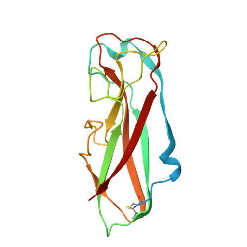
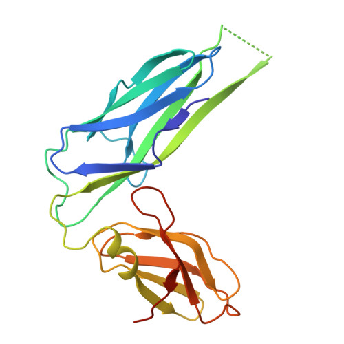
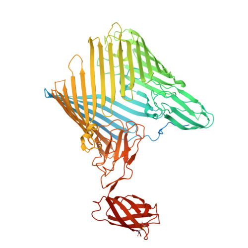
    Uropathogenic Escherichia coli strains use filamentous type 1 pili to adhere to and invade uroepithelial cells. The pilus consists of a flexible tip fibrillum, formed by the adhesin FimH and the subunits FimG and FimF. The pilus rod is a helical assembly of up to 3000 copies of the main subunit FimA, terminated by a single copy of the subunit FimI that anchors the rod to the assembly platform FimD in the outer membrane. Although type 1 pilus assembly can be completely reconstituted in vitro, the precise mechanism of assembly termination on FimD is still unknown. Here, we present cryo-electron microscopy structures of the fully assembled pilus with all its components prior to and after incorporation of FimI, capped with the assembly chaperone FimC. The structures reveal that FimD positions the proximal end of the pilus rod at an angle of ca. 50 degrees relative to the plane of the outer membrane. Specific interactions between FimI and FimC, absent in the equivalent FimA-FimC interface of the non-terminated pilus, stabilize the assembly-terminated state. In addition, we present structures of the transition region between the tip fibrillum and the helical rod, showing how FimF aligns the tip fibrillum along the rod axis.


  • Organizational Affiliation
    • Institute of Molecular Biology and Biophysics, ETH Zürich, Otto-Stern-Weg 5, Zürich, 8093, Switzerland.

Macromolecules
Find similar proteins by:  (by identity cutoff)  |  3D Structure
Entity ID: 1
MoleculeChains Sequence LengthOrganismDetailsImage
Fimbrin-like protein FimIA [auth B],
D [auth I]
160Escherichia coliMutation(s): 0 
Gene Names: fimIb4315JW5779
UniProt
Find proteins for P39264 (Escherichia coli (strain K12))
Explore P39264 
Go to UniProtKB:  P39264
Entity Groups  
Sequence Clusters30% Identity50% Identity70% Identity90% Identity95% Identity100% Identity
UniProt GroupP39264
Sequence Annotations
Expand
  • Reference Sequence
Find similar proteins by:  (by identity cutoff)  |  3D Structure
Entity ID: 2
MoleculeChains Sequence LengthOrganismDetailsImage
Chaperone protein FimCB [auth C]212Escherichia coliMutation(s): 0 
Gene Names: fimCb4316JW4279
UniProt
Find proteins for P31697 (Escherichia coli (strain K12))
Explore P31697 
Go to UniProtKB:  P31697
Entity Groups  
Sequence Clusters30% Identity50% Identity70% Identity90% Identity95% Identity100% Identity
UniProt GroupP31697
Sequence Annotations
Expand
  • Reference Sequence
Find similar proteins by:  (by identity cutoff)  |  3D Structure
Entity ID: 3
MoleculeChains Sequence LengthOrganismDetailsImage
Outer membrane usher protein FimDC [auth D]847Escherichia coliMutation(s): 0 
Gene Names: fimDb4317JW5780
UniProt
Find proteins for P30130 (Escherichia coli (strain K12))
Explore P30130 
Go to UniProtKB:  P30130
Entity Groups  
Sequence Clusters30% Identity50% Identity70% Identity90% Identity95% Identity100% Identity
UniProt GroupP30130
Sequence Annotations
Expand
  • Reference Sequence
Experimental Data & Validation

Experimental Data

  • Method: ELECTRON MICROSCOPY
  • Resolution: 4.20 Å
  • Aggregation State: PARTICLE 
  • Reconstruction Method: SINGLE PARTICLE 
EM Software:
TaskSoftware PackageVersion
MODEL REFINEMENTPHENIX1.21.1_5286
RECONSTRUCTIONcryoSPARC

Structure Validation

View Full Validation Report


Entry History & Funding Information

Deposition Data


Funding OrganizationLocationGrant Number
Swiss National Science FoundationSwitzerland310030_201234

Revision History  (Full details and data files)

  • Version 1.0: 2025-06-04
    Type: Initial release
  • Version 1.1: 2025-12-17
    Changes: Data collection, Database references